Copyright
©The Author(s) 2023.
World J Clin Cases. Apr 26, 2023; 11(12): 2780-2787
Published online Apr 26, 2023. doi: 10.12998/wjcc.v11.i12.2780
Published online Apr 26, 2023. doi: 10.12998/wjcc.v11.i12.2780
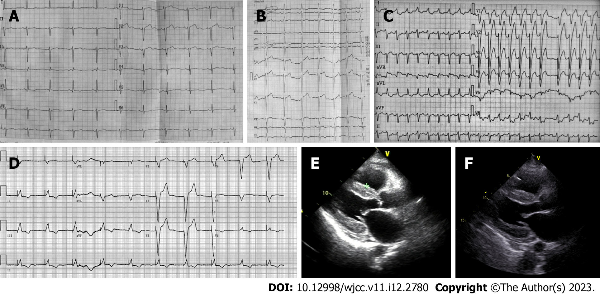
Figure 1 Serial changes on electrocardiography and echocardiography.
A: Electrocardiography (ECG) demonstrated a left anterior fascicular block, poor R-wave progression in leads V1-V3, and a non-specific T wave and ST-segment; B: 11 mo later, ECG demonstrated a new left bundle branch block, ST-segment depression in leads I and augmented unipolar limb lead (AVL) and T wave inversion in leads I, AVL, and V4-V6; C: 21 mo later, ECG demonstrated atrial fibrillation, left bundle branch block with wider QRS wave group, ST segment elevation in leads V1-V3, and T wave inversion in leads I, AVL and V4-V6; D: Echocardiography revealed characteristic sparkling and a granular texture in the ventricle wall; E: Pacing ECG was showed; F: Echocardiography revealed atrial enlargements and worsening systolic dysfunction (ejection fraction 48%).
- Citation: Gao M, Zhang WH, Zhang ZG, Yang N, Tong Q, Chen LP. Cardiac amyloidosis presenting as pulmonary arterial hypertension: A case report. World J Clin Cases 2023; 11(12): 2780-2787
- URL: https://www.wjgnet.com/2307-8960/full/v11/i12/2780.htm
- DOI: https://dx.doi.org/10.12998/wjcc.v11.i12.2780


