Copyright
©The Author(s) 2023.
World J Clin Cases. Apr 26, 2023; 11(12): 2637-2656
Published online Apr 26, 2023. doi: 10.12998/wjcc.v11.i12.2637
Published online Apr 26, 2023. doi: 10.12998/wjcc.v11.i12.2637
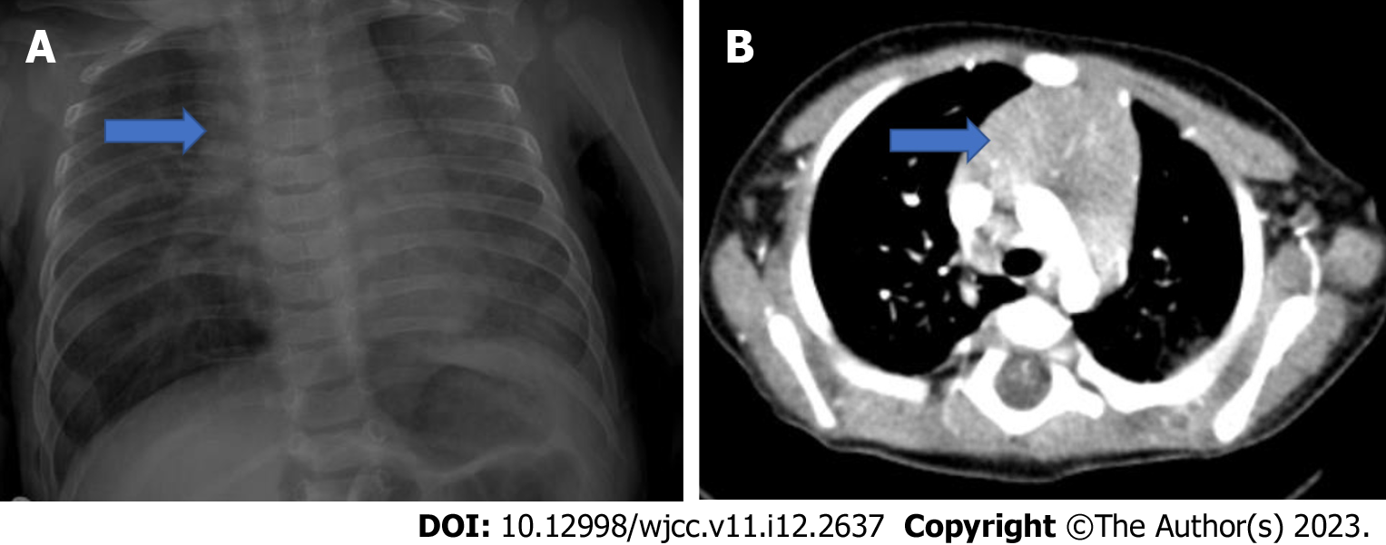
Figure 7 Normal thymus and axial computed tomography of a 4-mo-old patient.
A: Normal thymus on posteroanterior chest radiograph; B: Axial computed tomography (CT) examination of a 4-mo-old patient. In CT, the thymus (blue arrow) arcus is observed in the prevascular distance at the level of the aorta, its edges are convex, with homogeneous density and it does not cause a compression effect.
- Citation: Çinar HG, Gulmez AO, Üner Ç, Aydin S. Mediastinal lesions in children. World J Clin Cases 2023; 11(12): 2637-2656
- URL: https://www.wjgnet.com/2307-8960/full/v11/i12/2637.htm
- DOI: https://dx.doi.org/10.12998/wjcc.v11.i12.2637


