Copyright
©The Author(s) 2023.
World J Clin Cases. Apr 16, 2023; 11(11): 2549-2558
Published online Apr 16, 2023. doi: 10.12998/wjcc.v11.i11.2549
Published online Apr 16, 2023. doi: 10.12998/wjcc.v11.i11.2549
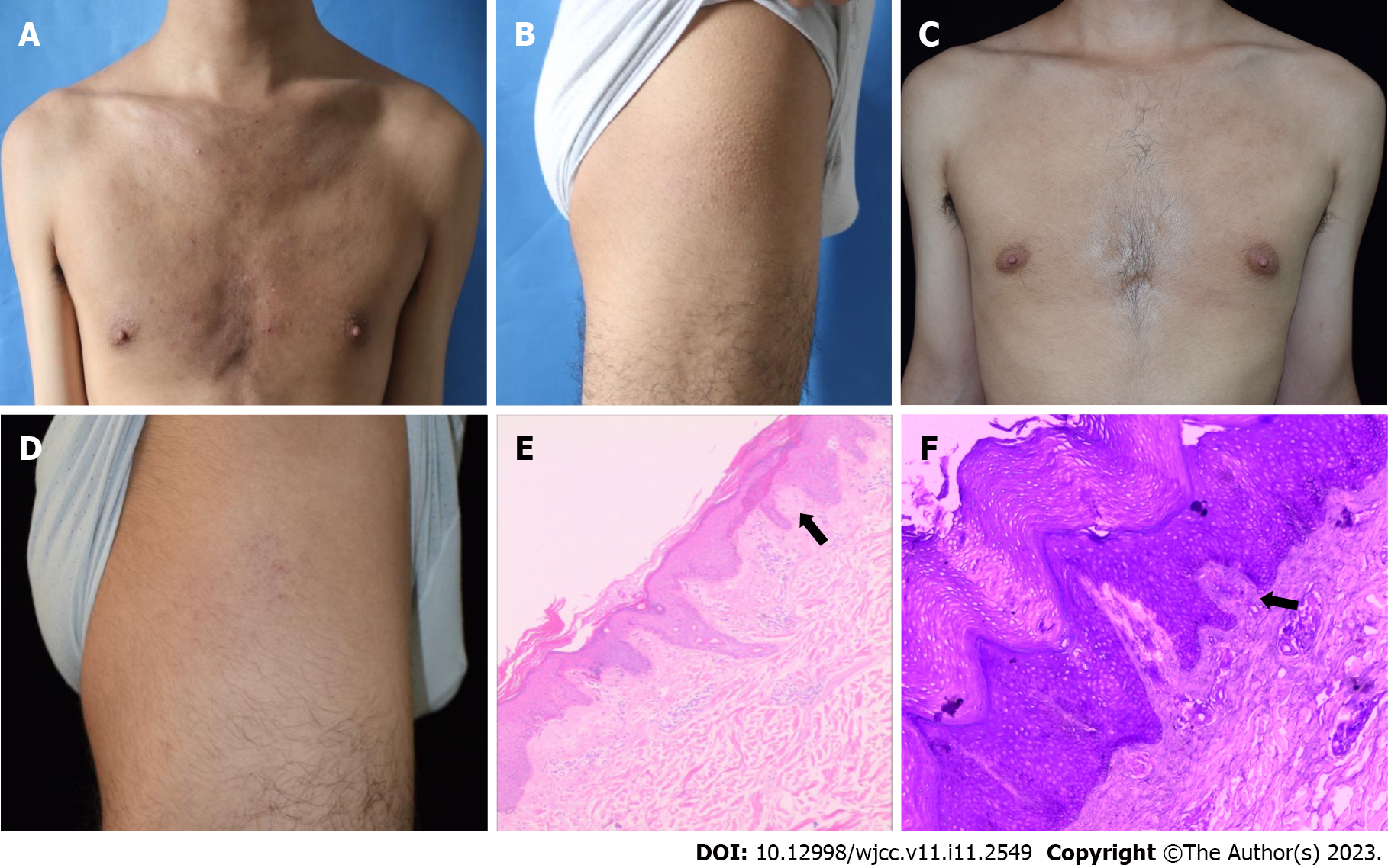
Figure 3 Case 3.
A and B: Dense distribution of hard brown papules was observed on the chest plus papules on the thighs, with a "goose bump" appearance presenting at baseline, suggestive of atopic dermatitis; C and D: After 16 wk of treatment, the rash on the lower limbs markedly improved, and the papules were reduced and became flat; E: Histopathology revealed hyperkeratosis with incomplete keratosis and irregular acanthosis. Further, a pink amorphous material can be seen in the dermal papilla (hematoxylin & eosin staining, × 100); F: Crystal violet staining was positive for amyloid (crystal violet staining, × 400). The arrow indicates amyloid deposits.
- Citation: Zhu Q, Gao BQ, Zhang JF, Shi LP, Zhang GQ. Successful treatment of lichen amyloidosis coexisting with atopic dermatitis by dupilumab: Four case reports. World J Clin Cases 2023; 11(11): 2549-2558
- URL: https://www.wjgnet.com/2307-8960/full/v11/i11/2549.htm
- DOI: https://dx.doi.org/10.12998/wjcc.v11.i11.2549


