Copyright
©The Author(s) 2023.
World J Clin Cases. Apr 16, 2023; 11(11): 2549-2558
Published online Apr 16, 2023. doi: 10.12998/wjcc.v11.i11.2549
Published online Apr 16, 2023. doi: 10.12998/wjcc.v11.i11.2549
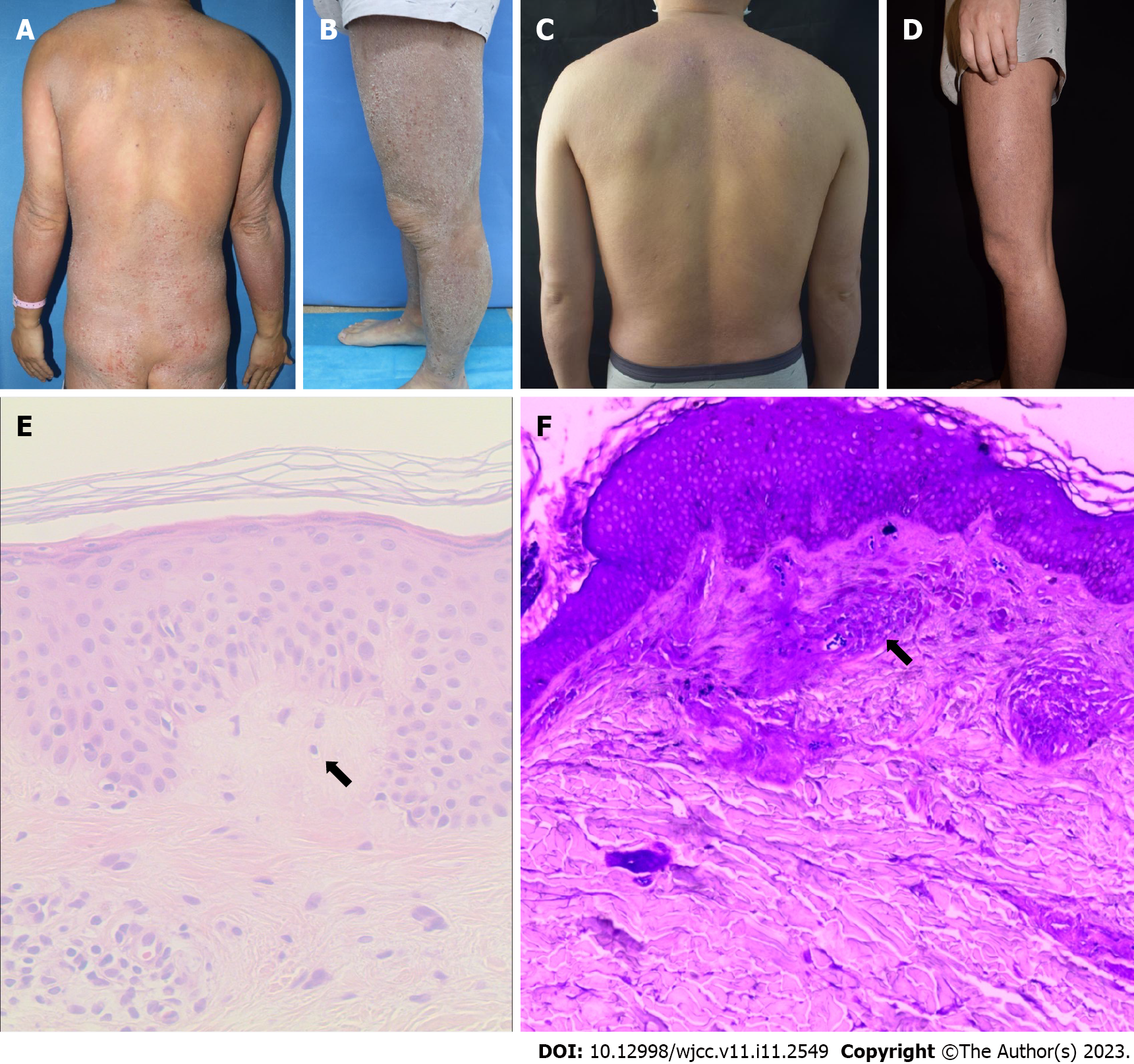
Figure 1 Case 1.
A and B: Dense hard brown plaques were seen on the trunk and limbs at baseline; C and D: After 16 wk of treatment with dupilumab, the trunk rash basically subsided, the lower limb rash markedly improved, and the plaques became flat. The remaining lesions on the thigh after treatment, showed a characteristic rippled appearance indicating lichen amyloidosis as a co-existing condition; E: Histopathological examination revealed mild hyperkeratosis of the epidermis, irregular hyperplasia of the spinous layer, and spongy edema, with the dermal papilla and upper dermis showing a uniformly reddish mass that expanded the dermal papillae and displaced the rete ridges laterally (hematoxylin & eosin staining, × 100); F: Crystal violet staining was positive for amyloid deposits in the dermal papillae and upper dermis with characteristic fissures (crystal violet staining, × 400). The arrow indicates amyloid deposits.
- Citation: Zhu Q, Gao BQ, Zhang JF, Shi LP, Zhang GQ. Successful treatment of lichen amyloidosis coexisting with atopic dermatitis by dupilumab: Four case reports. World J Clin Cases 2023; 11(11): 2549-2558
- URL: https://www.wjgnet.com/2307-8960/full/v11/i11/2549.htm
- DOI: https://dx.doi.org/10.12998/wjcc.v11.i11.2549


