Copyright
©The Author(s) 2023.
World J Clin Cases. Apr 16, 2023; 11(11): 2521-2527
Published online Apr 16, 2023. doi: 10.12998/wjcc.v11.i11.2521
Published online Apr 16, 2023. doi: 10.12998/wjcc.v11.i11.2521
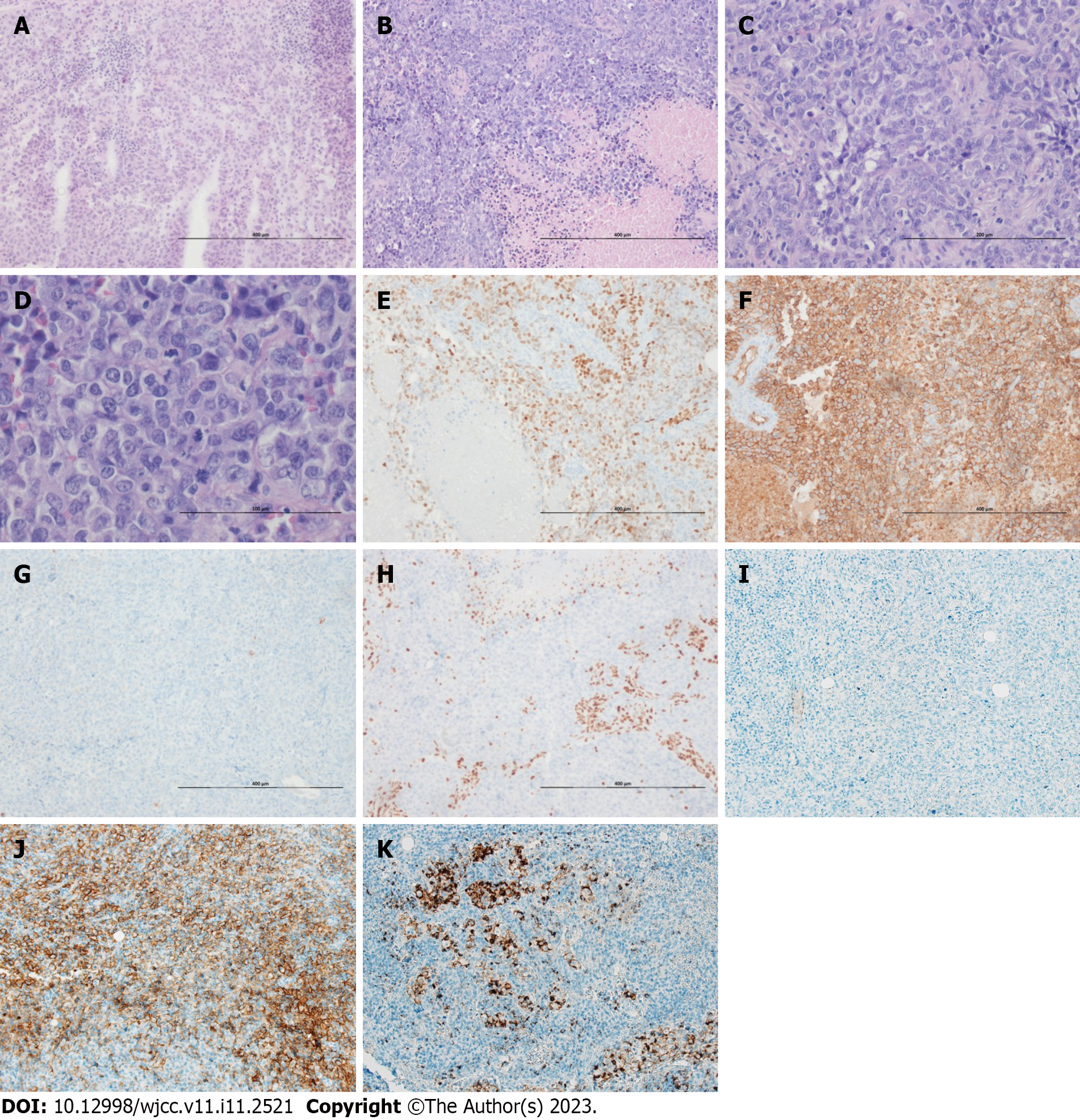
Figure 2 Histological features and immunostaining results of the tumor.
A and B: The frozen section shows a diffuse sheet of large round to epithelioid tumor cells on frozen section (A, × 100) and post fixation tissue section (B, × 100). Frequent necrotic foci are seen (B); C: The tumor cells show no glandular or squamous differentiation and exhibit moderate pleomorphism (× 200); D: The nuclei are monotonous and vesicular with conspicuous nucleoli. The tumor has eosinophilic cytoplasm and a markedly increased mitotic activity (× 400); E and F: Immunohistochemical staining for Spalt-like transcription factor 4 (SALL4) (E, × 100) and CD34 (F, × 100) exhibits positive results; G: The ancillary test, such as that for AE1/AE3 cytokeratin confirms the absence of epithelial differentiation (× 100); H: SMARCA4 staining exhibits diagnostic complete loss of expression (× 100); I: Anaplastic lymphoma kinase (ALK) (D5F3) shows negative activity (× 100); J and K: Programmed death-ligand 1 (PDL1) tests exhibit positivity (J, SP263, × 100; K, SP142, × 100). Staining method: Hematoxylin and eosin staining (A-D); polymer method (E-K).
- Citation: Kwon HJ, Jang MH. SMARCA4-deficient undifferentiated thoracic tumor: A case report. World J Clin Cases 2023; 11(11): 2521-2527
- URL: https://www.wjgnet.com/2307-8960/full/v11/i11/2521.htm
- DOI: https://dx.doi.org/10.12998/wjcc.v11.i11.2521


