Copyright
©The Author(s) 2023.
World J Clin Cases. Apr 16, 2023; 11(11): 2510-2520
Published online Apr 16, 2023. doi: 10.12998/wjcc.v11.i11.2510
Published online Apr 16, 2023. doi: 10.12998/wjcc.v11.i11.2510
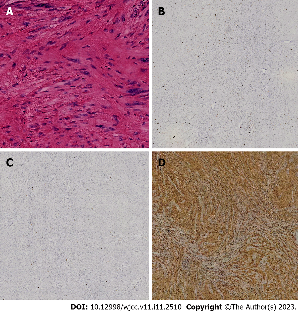
Figure 3 Histology and immunohistochemistry of the tumor.
A: Histopathologic findings revealed spindle-shaped cells in a fasciculated and disarrayed architecture, and no pathologic mitosis was observed (hematoxylin and eosin staining; (magnification, × 400); B: The mitotic activity rate was < 5% on Ki-67 staining (magnification, × 80); C: Immunochemical analysis revealed no staining with CD117 (magnification, × 80); D: Immunohistochemical examination revealed S-100 protein positivity (magnification, × 200).
- Citation: Mu YZ, Zhang Q, Zhao J, Liu Y, Kong LW, Ding ZX. Total removal of a large esophageal schwannoma by submucosal tunneling endoscopic resection: A case report and review of literature. World J Clin Cases 2023; 11(11): 2510-2520
- URL: https://www.wjgnet.com/2307-8960/full/v11/i11/2510.htm
- DOI: https://dx.doi.org/10.12998/wjcc.v11.i11.2510


