Copyright
©The Author(s) 2023.
World J Clin Cases. Apr 16, 2023; 11(11): 2510-2520
Published online Apr 16, 2023. doi: 10.12998/wjcc.v11.i11.2510
Published online Apr 16, 2023. doi: 10.12998/wjcc.v11.i11.2510
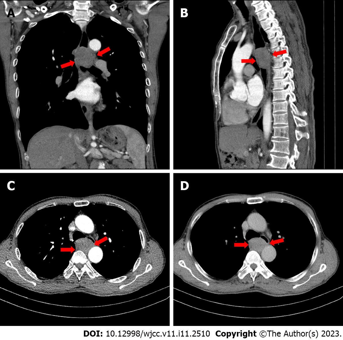
Figure 1 Plain and contrast-enhanced chest computed tomography.
A: Coronal view of chest computed tomography (CT) showed that the tumor in the middle and upper esophagus had clear boundaries and homogeneous density; B: Sagittal view of the CT scan revealed the mass was located in the posterior mediastinum, and the upper and lower diameters were larger than the anterior and posterior diameters; C and D: Axial view of CT demonstrated the tumor presented homogeneous enhancement.
- Citation: Mu YZ, Zhang Q, Zhao J, Liu Y, Kong LW, Ding ZX. Total removal of a large esophageal schwannoma by submucosal tunneling endoscopic resection: A case report and review of literature. World J Clin Cases 2023; 11(11): 2510-2520
- URL: https://www.wjgnet.com/2307-8960/full/v11/i11/2510.htm
- DOI: https://dx.doi.org/10.12998/wjcc.v11.i11.2510


