Copyright
©The Author(s) 2023.
World J Clin Cases. Apr 16, 2023; 11(11): 2464-2473
Published online Apr 16, 2023. doi: 10.12998/wjcc.v11.i11.2464
Published online Apr 16, 2023. doi: 10.12998/wjcc.v11.i11.2464
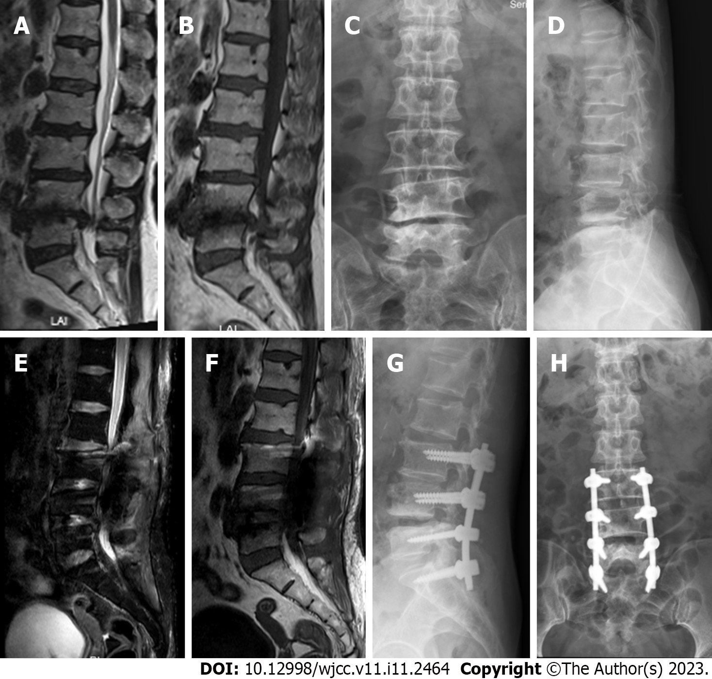
Figure 3 Magnetic resonance imaging and X-ray findings of the first patient.
A–D: Preoperative images showing lumbar disc herniation and lumbar spinal stenosis at L3–S1 level; E–H: Images at 1 wk after surgery showing the lack of clear signs of cerebrospinal fluid leakage or pseudocyst formation.
- Citation: Xu C, Dong RP, Cheng XL, Zhao JW. Late presentation of dural tears: Two case reports and review of literature. World J Clin Cases 2023; 11(11): 2464-2473
- URL: https://www.wjgnet.com/2307-8960/full/v11/i11/2464.htm
- DOI: https://dx.doi.org/10.12998/wjcc.v11.i11.2464


