Copyright
©The Author(s) 2023.
World J Clin Cases. Apr 6, 2023; 11(10): 2329-2335
Published online Apr 6, 2023. doi: 10.12998/wjcc.v11.i10.2329
Published online Apr 6, 2023. doi: 10.12998/wjcc.v11.i10.2329
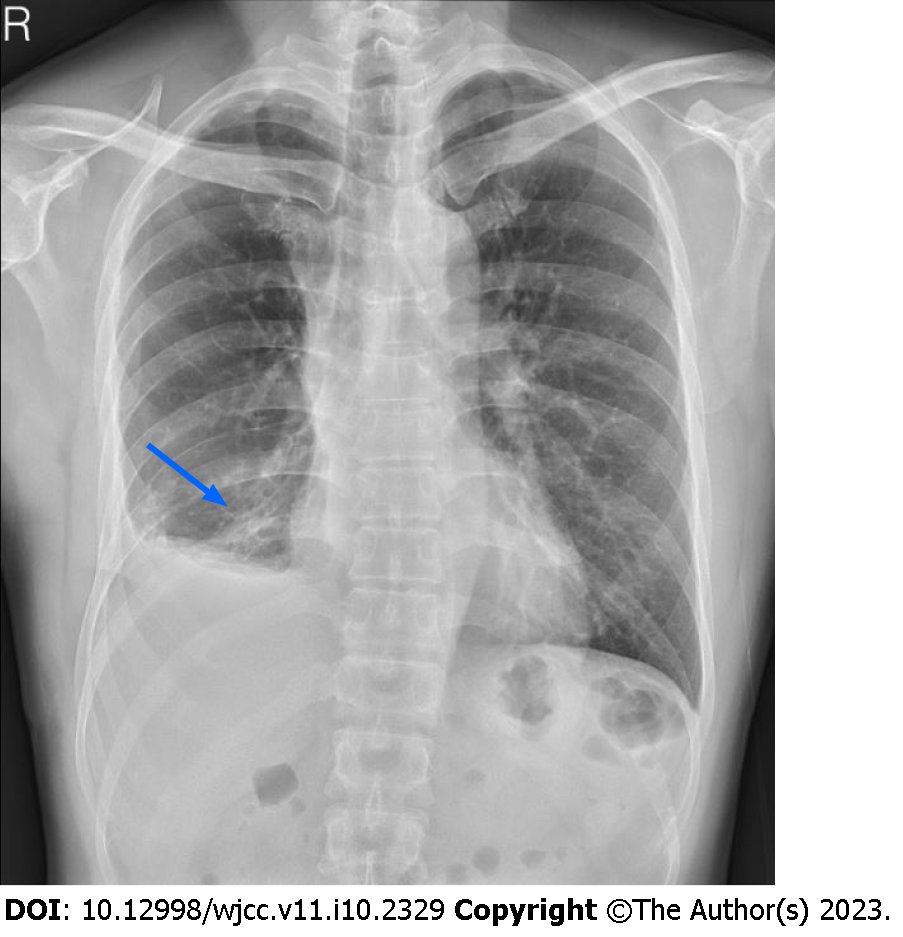
Figure 4 Follow-up chest radiograph obtained two years postoperatively.
The image shows calcified shadows over the right lower lung field (blue arrow).
- Citation: Hsu FC, Huang TW, Pu TW. Formation of a rare curve-shaped thoracolith documented on serial chest computed tomography images: A case report. World J Clin Cases 2023; 11(10): 2329-2335
- URL: https://www.wjgnet.com/2307-8960/full/v11/i10/2329.htm
- DOI: https://dx.doi.org/10.12998/wjcc.v11.i10.2329


