Copyright
©The Author(s) 2023.
World J Clin Cases. Apr 6, 2023; 11(10): 2329-2335
Published online Apr 6, 2023. doi: 10.12998/wjcc.v11.i10.2329
Published online Apr 6, 2023. doi: 10.12998/wjcc.v11.i10.2329
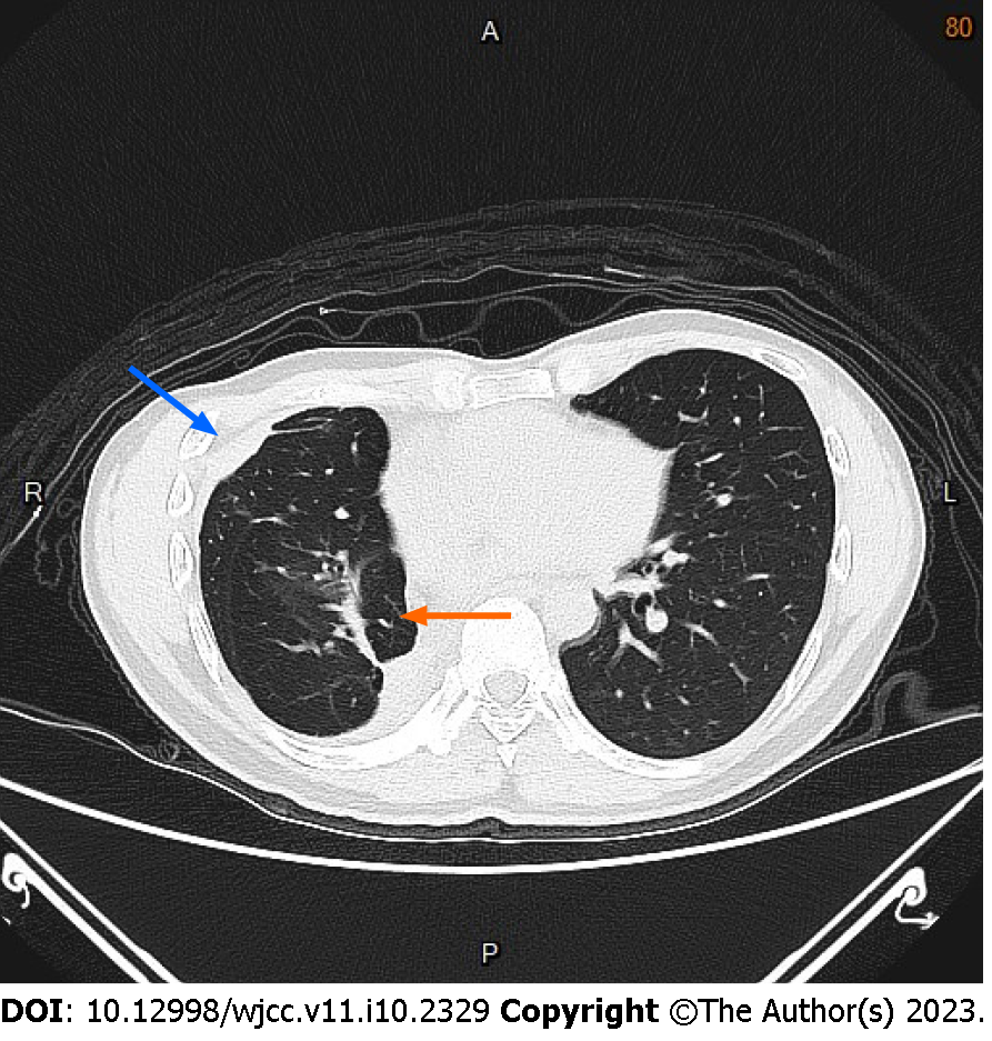
Figure 3 Follow-up chest computed tomography image obtained two years after the initial therapy.
Axial image showing thickening of the right pleura (blue arrow) and focal atelectasis (orange arrow) in the right middle lung lobe.
- Citation: Hsu FC, Huang TW, Pu TW. Formation of a rare curve-shaped thoracolith documented on serial chest computed tomography images: A case report. World J Clin Cases 2023; 11(10): 2329-2335
- URL: https://www.wjgnet.com/2307-8960/full/v11/i10/2329.htm
- DOI: https://dx.doi.org/10.12998/wjcc.v11.i10.2329


