Copyright
©The Author(s) 2023.
World J Clin Cases. Apr 6, 2023; 11(10): 2321-2328
Published online Apr 6, 2023. doi: 10.12998/wjcc.v11.i10.2321
Published online Apr 6, 2023. doi: 10.12998/wjcc.v11.i10.2321
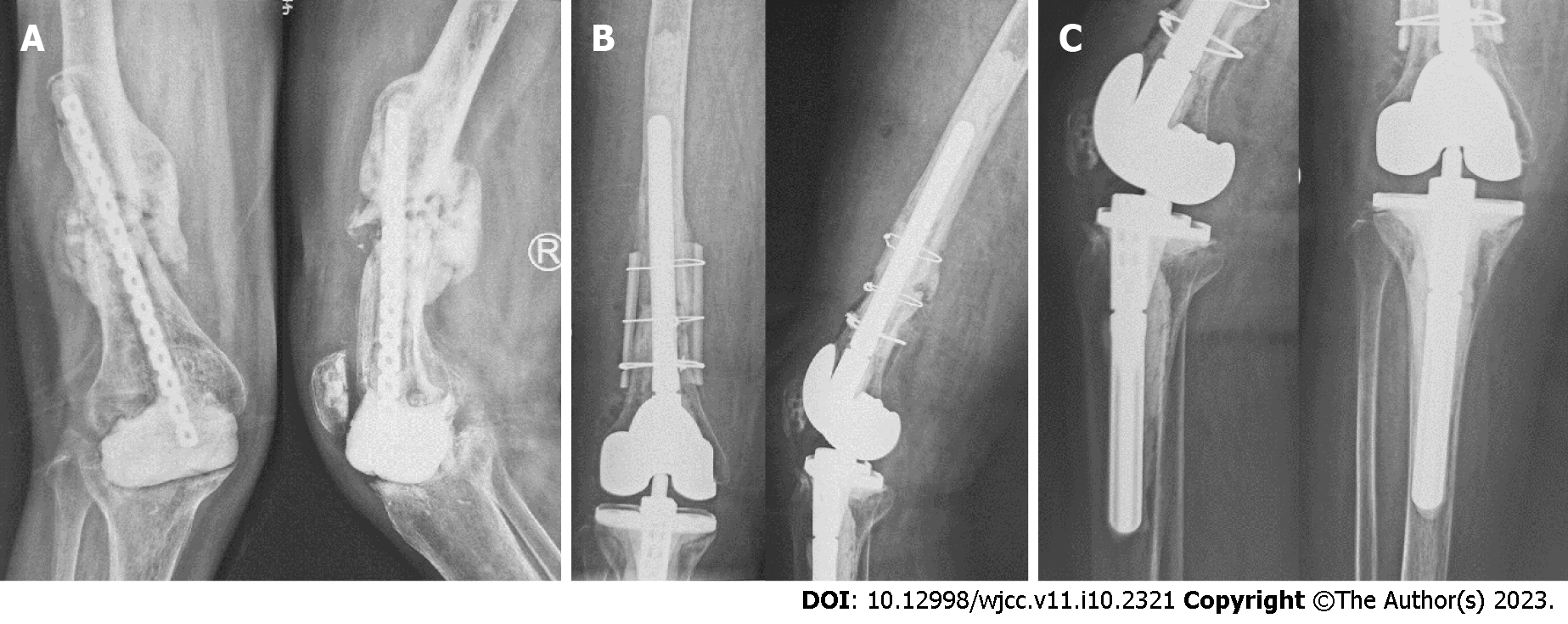
Figure 3 X-ray images.
A: Anterior–posterior and lateral X-ray images of the right knee before the second stage operation showing that the cement spacer slightly displaced to the medial side, the proximal end of internal fixator came out through the middle femoral shaft, callus formed at the previous fracture of the femur but not healing yet, and bone defects existed in the distal femur and proximal tibia, which resulted in valgus deformity of the right knee (May 2020); B and C: Anterior–posterior and lateral X-ray images of the right knee after the second stage operation showing no loosening of cemented revision knee prosthesis and that the femoral fracture was fixed with intramedullary stem and extramedullary cortical splints (May 2020).
- Citation: Hao LJ, Wen PF, Zhang YM, Song W, Chen J, Ma T. Treatment of periprosthetic knee infection and coexistent periprosthetic fracture: A case report and literature review. World J Clin Cases 2023; 11(10): 2321-2328
- URL: https://www.wjgnet.com/2307-8960/full/v11/i10/2321.htm
- DOI: https://dx.doi.org/10.12998/wjcc.v11.i10.2321


