Copyright
©The Author(s) 2023.
World J Clin Cases. Apr 6, 2023; 11(10): 2321-2328
Published online Apr 6, 2023. doi: 10.12998/wjcc.v11.i10.2321
Published online Apr 6, 2023. doi: 10.12998/wjcc.v11.i10.2321
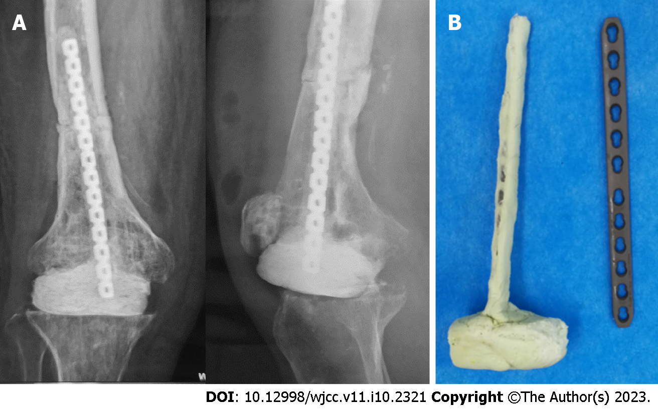
Figure 2 X-ray and intraoperative images.
A: Anterior–posterior and lateral X-ray images of the right knee after the first stage operation showing favorable position of the cement spacer and satisfactory reduction and fixation of the femoral fracture (July 2019); B: A hand-made T-shaped antibiotic-laden cement spacer for knee gap occupation and femoral fracture fixation.
- Citation: Hao LJ, Wen PF, Zhang YM, Song W, Chen J, Ma T. Treatment of periprosthetic knee infection and coexistent periprosthetic fracture: A case report and literature review. World J Clin Cases 2023; 11(10): 2321-2328
- URL: https://www.wjgnet.com/2307-8960/full/v11/i10/2321.htm
- DOI: https://dx.doi.org/10.12998/wjcc.v11.i10.2321


