Copyright
©The Author(s) 2023.
World J Clin Cases. Apr 6, 2023; 11(10): 2321-2328
Published online Apr 6, 2023. doi: 10.12998/wjcc.v11.i10.2321
Published online Apr 6, 2023. doi: 10.12998/wjcc.v11.i10.2321
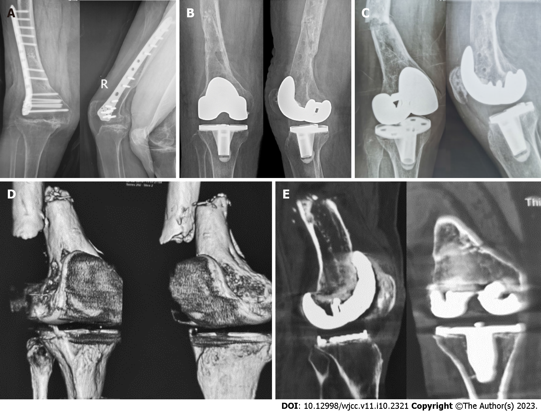
Figure 1 X-ray and computed tomography images.
A: Anterior–posterior and lateral X-ray images of the right knee before primary total knee arthroplasty showing an end-stage osteoarthritis of the right knee with valgus deformity and a hardware fixed on right femur without signs of fracture (December 2018); B: Anterior–posterior and lateral X-ray images of the right knee after the primary total knee arthroplasty showing satisfactory position of the knee prosthesis and that the original hardware was completely removed without fracture (December 2018); C: Anterior–posterior and lateral X-ray images of the right knee after fall showing the distal femur fracture with significant displacement and that the knee prosthesis was seemingly stable (July 2019); D: Three-dimensional computed tomography (CT) images showing fractures of the right distal femur with severe displacement (July 2019); E: Coronal and sagittal CT images showing radiolucent lines around the femoral and tibial prosthesis (July 2019).
- Citation: Hao LJ, Wen PF, Zhang YM, Song W, Chen J, Ma T. Treatment of periprosthetic knee infection and coexistent periprosthetic fracture: A case report and literature review. World J Clin Cases 2023; 11(10): 2321-2328
- URL: https://www.wjgnet.com/2307-8960/full/v11/i10/2321.htm
- DOI: https://dx.doi.org/10.12998/wjcc.v11.i10.2321


