Copyright
©The Author(s) 2023.
World J Clin Cases. Apr 6, 2023; 11(10): 2282-2289
Published online Apr 6, 2023. doi: 10.12998/wjcc.v11.i10.2282
Published online Apr 6, 2023. doi: 10.12998/wjcc.v11.i10.2282
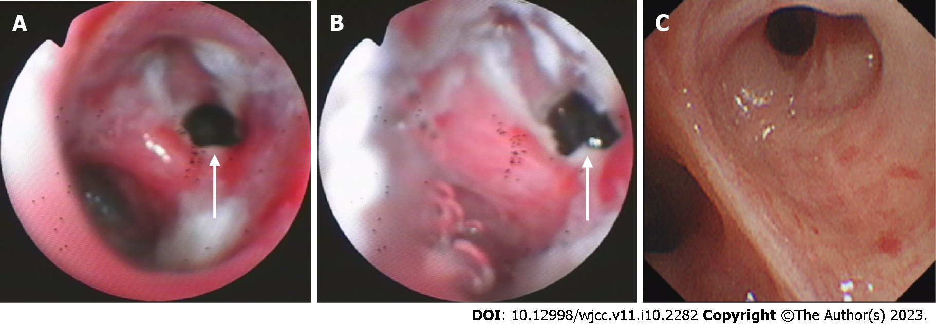
Figure 2 Bronchoscopic view of the right upper bronchus.
A: Bronchoscopy at admission shows a 5 mm fistula (white arrows) of the right upper bronchus, along with erythema, erosion, and hyperplasia of the apical bronchus, and a small amount of necrotic material; B: Bronchoscopy is performed 1 mo after the anti-tuberculosis treatment; C: Bronchoscopy is performed 6 mo after the anti-tuberculosis treatment.
- Citation: Shen L, Jiang YH, Dai XY. Successful surgical treatment of bronchopleural fistula caused by severe pulmonary tuberculosis: A case report and review of literature. World J Clin Cases 2023; 11(10): 2282-2289
- URL: https://www.wjgnet.com/2307-8960/full/v11/i10/2282.htm
- DOI: https://dx.doi.org/10.12998/wjcc.v11.i10.2282


