Copyright
©The Author(s) 2023.
World J Clin Cases. Jan 6, 2023; 11(1): 242-248
Published online Jan 6, 2023. doi: 10.12998/wjcc.v11.i1.242
Published online Jan 6, 2023. doi: 10.12998/wjcc.v11.i1.242
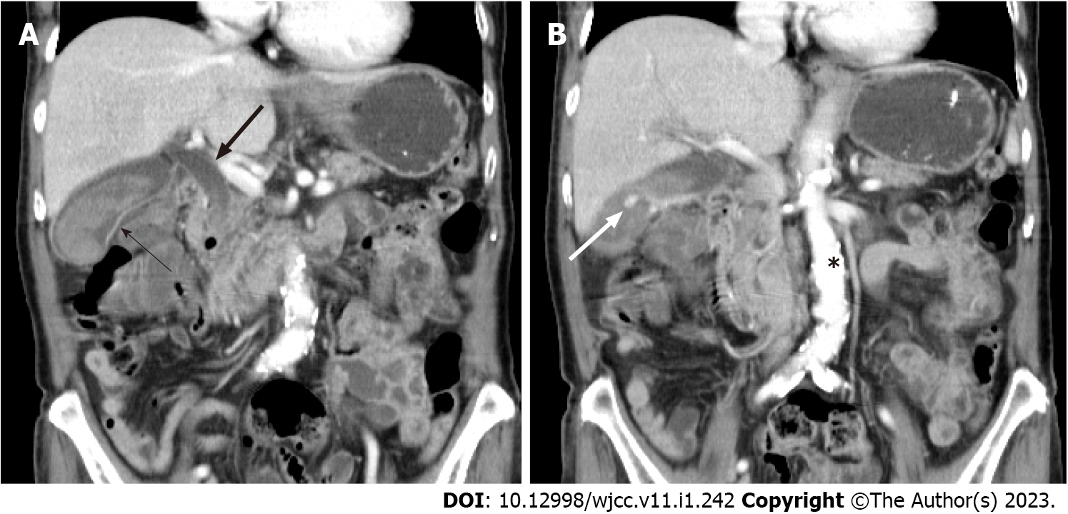
Figure 2 Sagittal views of contrast-enhanced abdominal computed tomography.
A: A thin-walled gallbladder with wall enhancement (thin arrow) with common bile duct dilatation (thick arrow); B: A hyperattenuated pseudoaneurysm (white arrow) and atherosclerotic aorta (*).
- Citation: Liu YL, Hsieh CT, Yeh YJ, Liu H. Cystic artery pseudoaneurysm: A case report. World J Clin Cases 2023; 11(1): 242-248
- URL: https://www.wjgnet.com/2307-8960/full/v11/i1/242.htm
- DOI: https://dx.doi.org/10.12998/wjcc.v11.i1.242


