Copyright
©The Author(s) 2023.
World J Clin Cases. Jan 6, 2023; 11(1): 233-241
Published online Jan 6, 2023. doi: 10.12998/wjcc.v11.i1.233
Published online Jan 6, 2023. doi: 10.12998/wjcc.v11.i1.233
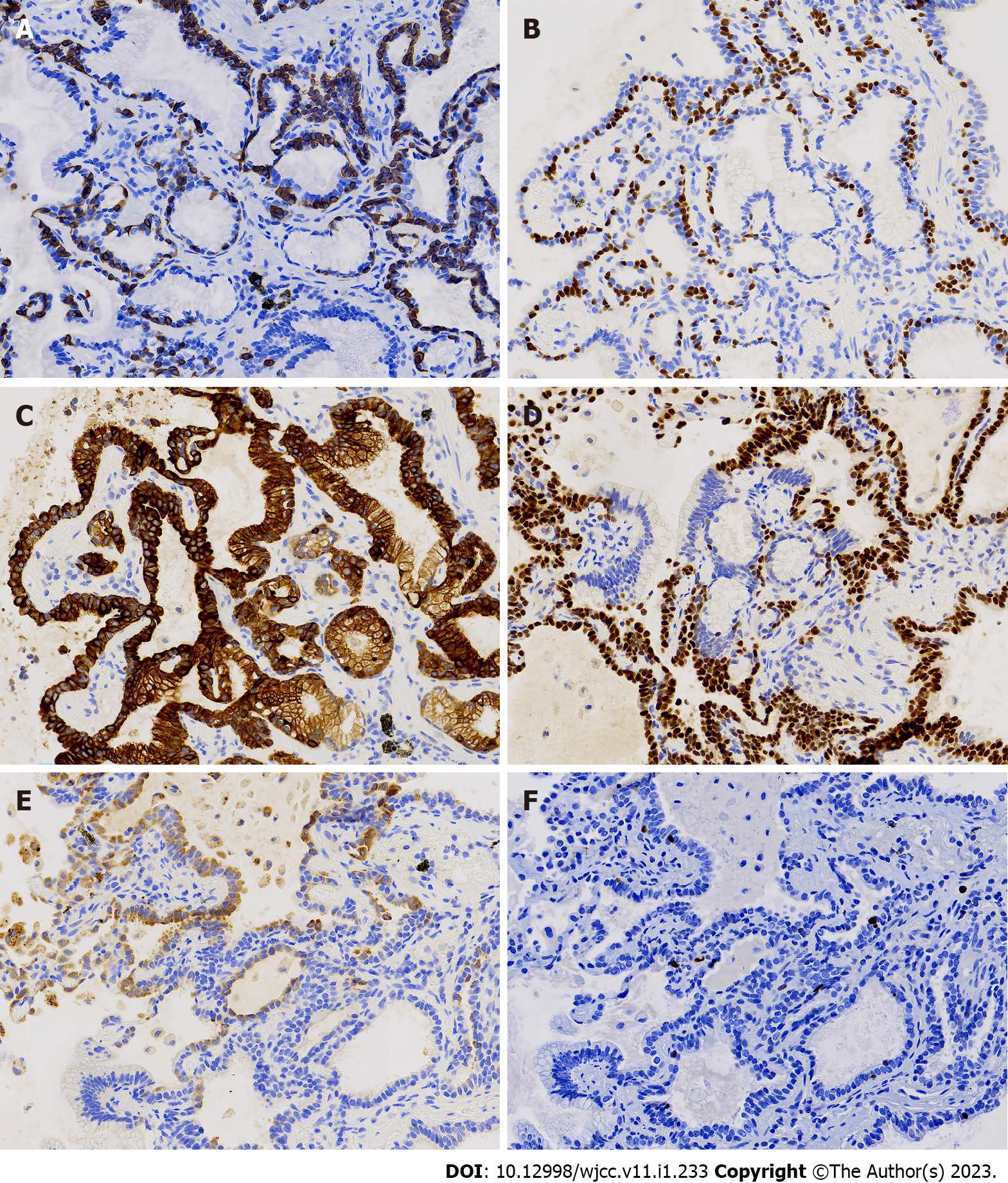
Figure 4 Immunohistochemical staining of the bronchiole adenoma area.
A: CK5/6 (×400); B: p40 (×400); C: CK7 (×400); D: TTF-1 (×400); E: Napsin A (×400); F: Ki-67 (×400). CK5/6 and p40 were expressed continuously in the basal cell layer in some areas, and had a discontinuous skipping pattern in other areas. CK7 had diffuse expression in basal cells and luminal cells. TTF-1 was expressed in basal cells, ciliated cells, and cubic cells, but not mucinous cells. Luminal cells had a patchy weak expression of napsin A. The Ki-67 index was low.
- Citation: Liu XL, Miao CF, Li M, Li P. Malignant transformation of pulmonary bronchiolar adenoma into mucinous adenocarcinoma: A case report. World J Clin Cases 2023; 11(1): 233-241
- URL: https://www.wjgnet.com/2307-8960/full/v11/i1/233.htm
- DOI: https://dx.doi.org/10.12998/wjcc.v11.i1.233


