Copyright
©The Author(s) 2023.
World J Clin Cases. Jan 6, 2023; 11(1): 233-241
Published online Jan 6, 2023. doi: 10.12998/wjcc.v11.i1.233
Published online Jan 6, 2023. doi: 10.12998/wjcc.v11.i1.233
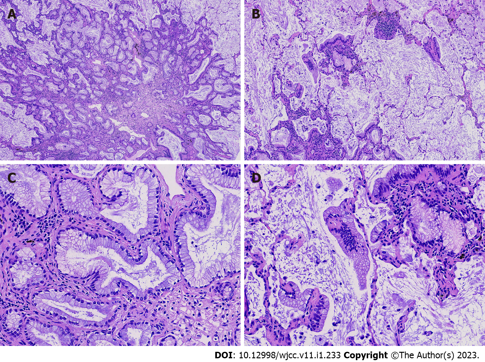
Figure 3 Hematoxylin and eosin staining of the mucinous adenocarcinoma area.
A: The tumor was arranged in a glandular structure (×40); B and C: There was a skipping growth pattern around the tissues (B: ×200, and C: ×400); D: It consisted of columnar cells without a clear basal cell layer (×400).
- Citation: Liu XL, Miao CF, Li M, Li P. Malignant transformation of pulmonary bronchiolar adenoma into mucinous adenocarcinoma: A case report. World J Clin Cases 2023; 11(1): 233-241
- URL: https://www.wjgnet.com/2307-8960/full/v11/i1/233.htm
- DOI: https://dx.doi.org/10.12998/wjcc.v11.i1.233


