Copyright
©The Author(s) 2023.
World J Clin Cases. Jan 6, 2023; 11(1): 233-241
Published online Jan 6, 2023. doi: 10.12998/wjcc.v11.i1.233
Published online Jan 6, 2023. doi: 10.12998/wjcc.v11.i1.233
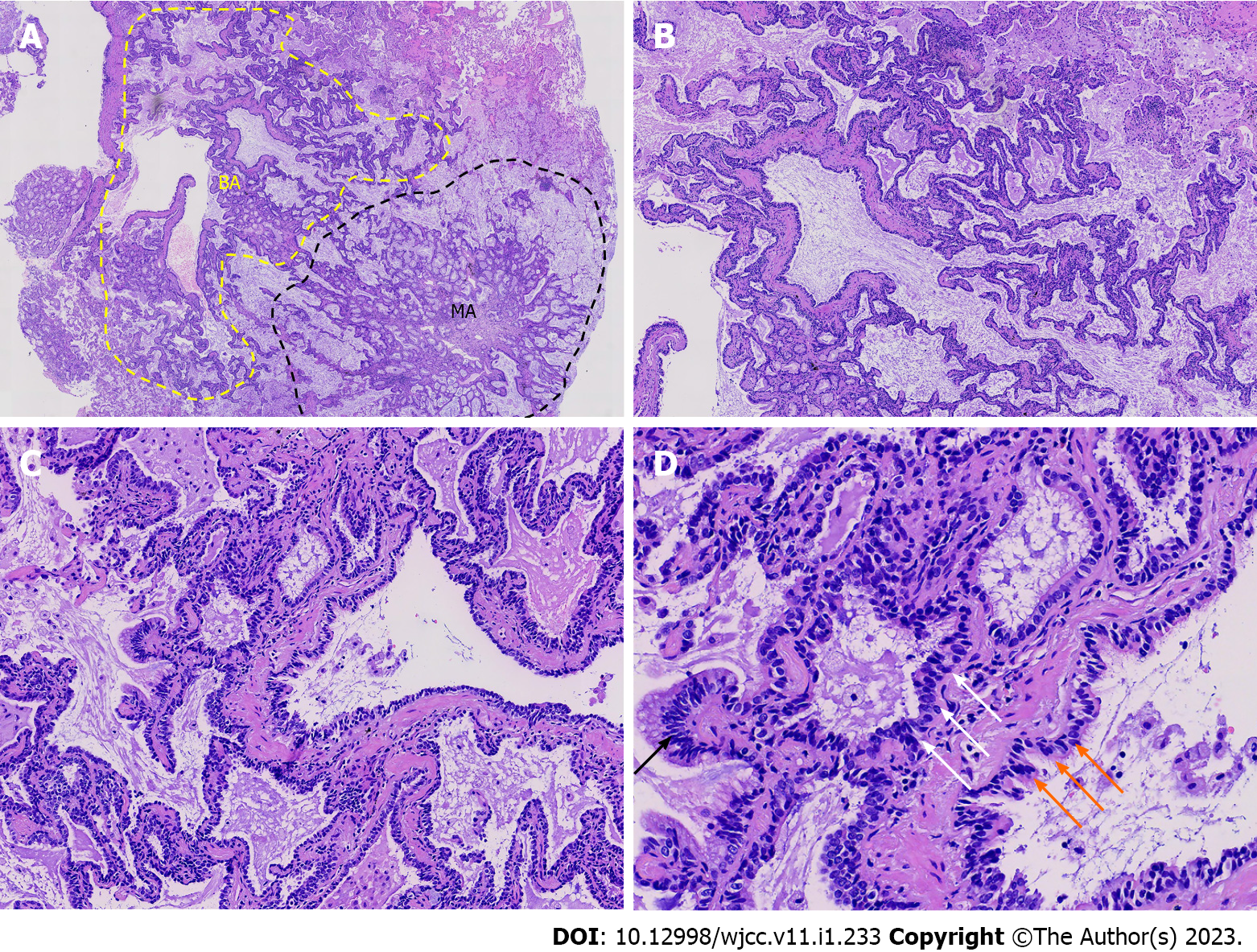
Figure 2 Hematoxylin and eosin staining of the whole tumor.
A: The tumor consisted of two general areas (×100): A bronchiole adenoma (BA) (yellow dotted line) area and a mucinous adenocarcinoma (black dotted line) area; B and C: The BA area consisted of a bilayered structure of luminal cells and basal cells that were arranged in glandular, papillary, and flat structures (B: ×100, and C: ×200); D: At high magnification (×400), basal cells (white arrows), ciliated cells (orange arrows) or cubic/Low columnar cells and locally abundant mucinous cells (black arrow), without significant atypia or pathological mitosis, were evident in the luminal epithelium. BA: Bronchiole adenoma; MA: Mucinous adenocarcinoma.
- Citation: Liu XL, Miao CF, Li M, Li P. Malignant transformation of pulmonary bronchiolar adenoma into mucinous adenocarcinoma: A case report. World J Clin Cases 2023; 11(1): 233-241
- URL: https://www.wjgnet.com/2307-8960/full/v11/i1/233.htm
- DOI: https://dx.doi.org/10.12998/wjcc.v11.i1.233


