Copyright
©The Author(s) 2023.
World J Clin Cases. Jan 6, 2023; 11(1): 164-171
Published online Jan 6, 2023. doi: 10.12998/wjcc.v11.i1.164
Published online Jan 6, 2023. doi: 10.12998/wjcc.v11.i1.164
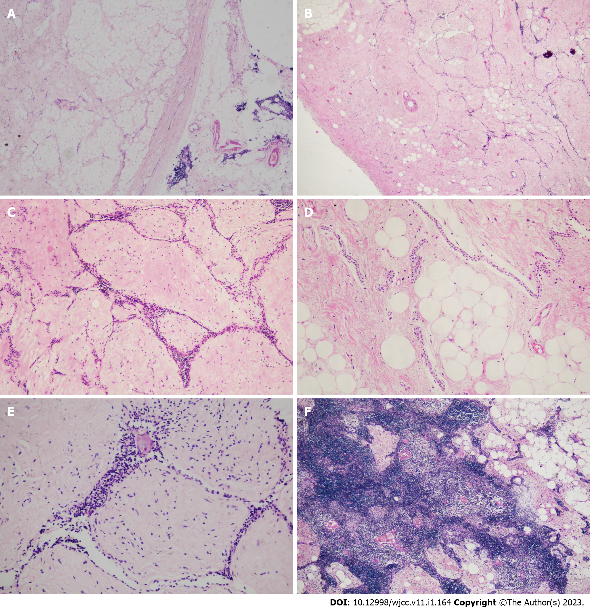
Figure 2 Histological features of the thymic lipofibroadenoma.
A: The tumor was all well circumscribed, and a clear connective capsule was observed between the tumor and the remaining thymus. [hematoxylin and eosin (H&E) staining, × 40]; B and C: The tumor was composed of fibrotic and hyaline stroma, narrow strands of epithelial cells and adipose tissues. The blank-looking epithelial cells formed narrow cords and interspersed in fibrotic and hyaline stroma. (H&E staining, B, × 40; C, × 100); D: Some adipocytes were mixed with the fibrotic stroma. Abundant adipose tissue also can be seen in some areas. (H&E staining, × 100); E: A few lymphocytes were mixed with the epithelial cells. Residual Hassall corpuscles and small calcifications could be seen in some region. (H&E staining, × 200); F: In case 2, hyperplastic thymic tissue was observed in part of the tumor. (H&E staining, × 100).
- Citation: Yang MQ, Wang ZQ, Chen LQ, Gao SM, Fu XN, Zhang HN, Zhang KX, Xu HT. Thymic lipofibroadenomas: Three case reports. World J Clin Cases 2023; 11(1): 164-171
- URL: https://www.wjgnet.com/2307-8960/full/v11/i1/164.htm
- DOI: https://dx.doi.org/10.12998/wjcc.v11.i1.164


