Copyright
©The Author(s) 2023.
World J Clin Cases. Jan 6, 2023; 11(1): 135-142
Published online Jan 6, 2023. doi: 10.12998/wjcc.v11.i1.135
Published online Jan 6, 2023. doi: 10.12998/wjcc.v11.i1.135
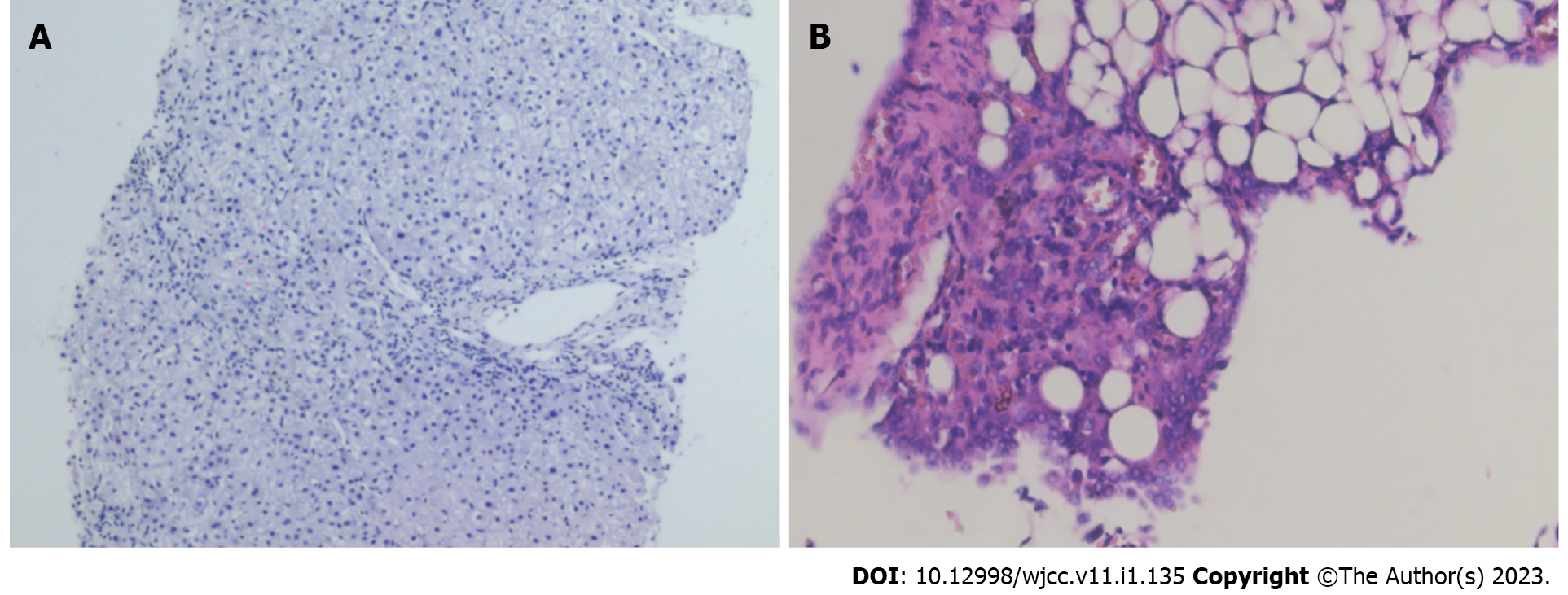
Figure 2 Pathology results.
A: The liver biopsy. Punctate necrosis and old bridging necrosis in the liver parenchyma and a small amount of eosinophilic infiltration in the portal area; B: The peritoneal biopsy. Inflammatory cell infiltration in fibro-adipose tissue and proliferation of small blood vessels, indicating inflammatory lesions.
- Citation: Zhou XL, Chang YH, Li L, Ren J, Wu XL, Zhang X, Wu P, Tang SH. Polyneuropathy organomegaly endocrinopathy M-protein and skin changes syndrome with ascites as an early-stage manifestation: A case report. World J Clin Cases 2023; 11(1): 135-142
- URL: https://www.wjgnet.com/2307-8960/full/v11/i1/135.htm
- DOI: https://dx.doi.org/10.12998/wjcc.v11.i1.135


