Copyright
©The Author(s) 2022.
World J Clin Cases. Mar 26, 2022; 10(9): 2961-2968
Published online Mar 26, 2022. doi: 10.12998/wjcc.v10.i9.2961
Published online Mar 26, 2022. doi: 10.12998/wjcc.v10.i9.2961
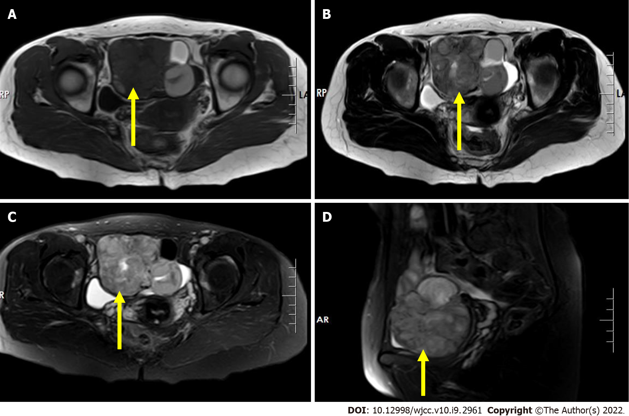
Figure 2 Pre-operative pelvis magnetic resonance imaging scan.
A: T1-weighted image shows an irregular mass with mixed signal in the right adnexal region with iso/hypo-signal intensity (axial, yellow arrow); B: T2-weighted image shows an irregular mass with a heterogeneous slightly high signal intensity in the right adnexal region (axial, yellow arrow); C and D: Short time of inversion recovery image shows an irregular mass with a heterogeneous slightly high signal intensity in the right adnexal region (yellow arrow) (C: Cornal; D: Sagittal).
- Citation: Xiao W, Zhou JR, Chen D. Malignant struma ovarii with papillary carcinoma combined with retroperitoneal lymph node metastasis: A case report. World J Clin Cases 2022; 10(9): 2961-2968
- URL: https://www.wjgnet.com/2307-8960/full/v10/i9/2961.htm
- DOI: https://dx.doi.org/10.12998/wjcc.v10.i9.2961


