Copyright
©The Author(s) 2022.
World J Clin Cases. Mar 26, 2022; 10(9): 2961-2968
Published online Mar 26, 2022. doi: 10.12998/wjcc.v10.i9.2961
Published online Mar 26, 2022. doi: 10.12998/wjcc.v10.i9.2961
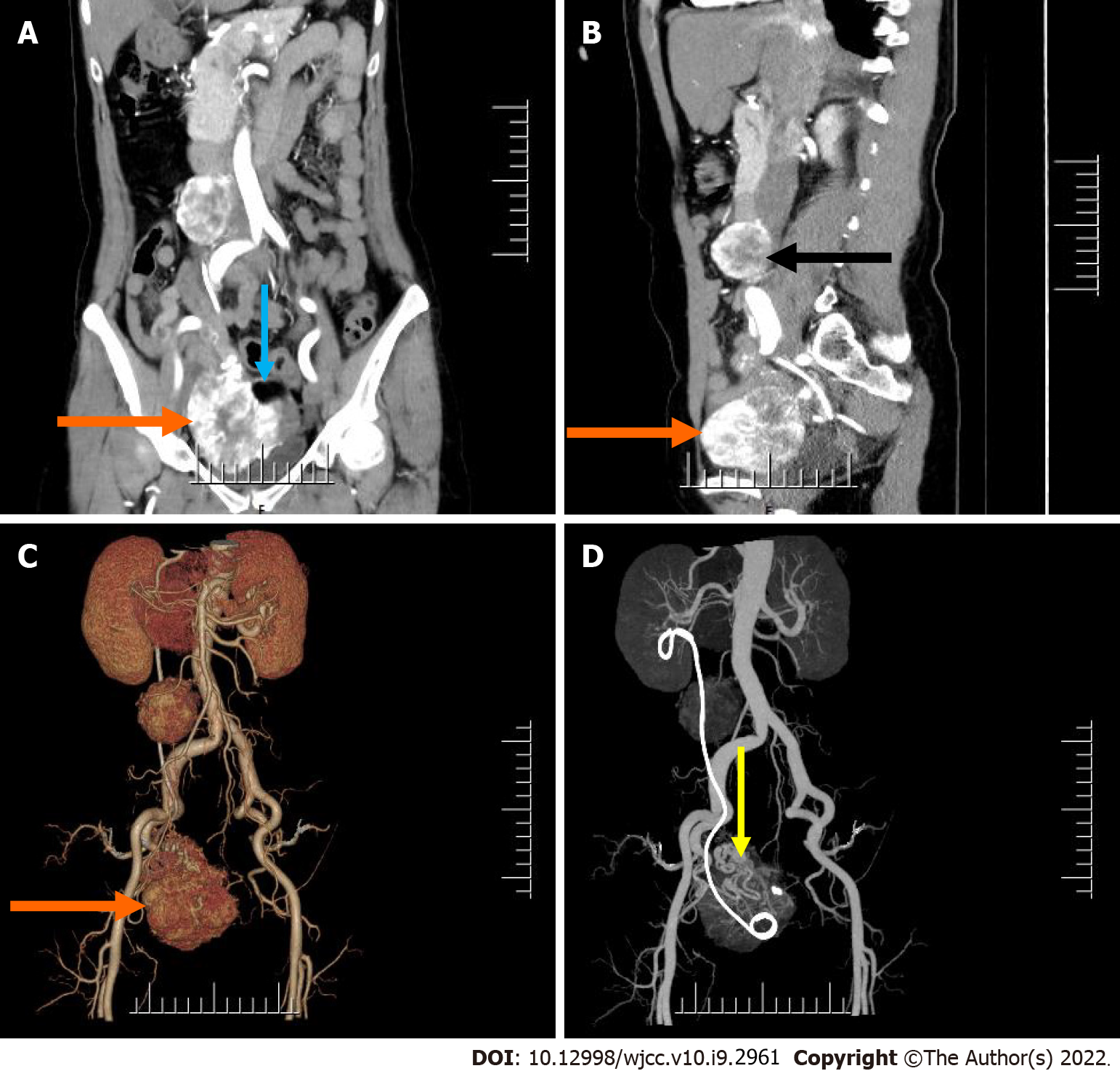
Figure 1 Pre-operative abdomen computed tomography contrast-enhanced scan.
A and B: Computed tomography (CT) images showed an irregular mass in the right adnexal region (orange arrow) and a round mass in the right retroperitoneum (black arrow) (A: Cornal; B: Sagittal); C and D: Post-processed CT images showed 3D reconstruction of both lesions (orange and yellow arrow).
- Citation: Xiao W, Zhou JR, Chen D. Malignant struma ovarii with papillary carcinoma combined with retroperitoneal lymph node metastasis: A case report. World J Clin Cases 2022; 10(9): 2961-2968
- URL: https://www.wjgnet.com/2307-8960/full/v10/i9/2961.htm
- DOI: https://dx.doi.org/10.12998/wjcc.v10.i9.2961


