Copyright
©The Author(s) 2022.
World J Clin Cases. Mar 26, 2022; 10(9): 2954-2960
Published online Mar 26, 2022. doi: 10.12998/wjcc.v10.i9.2954
Published online Mar 26, 2022. doi: 10.12998/wjcc.v10.i9.2954
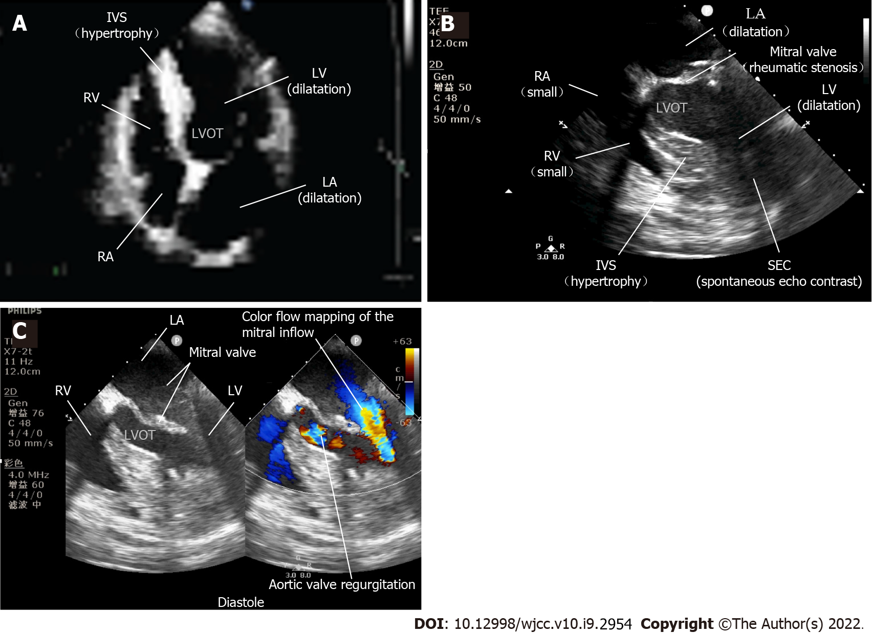
Figure 1 Perioperative echocardiography.
A: Preoperative transthoracic echocardiography (TTE) showed rheumatic heart disease; B: TEE revealed that the cardiac motion ceased. The right cardiac chambers were small, the rheumatic mitral stenosis and spontaneous echo contrast indicated blood stasis in the left cardiac chambers; C: TEE assisting with the weaning of percutaneous cardiopulmonary bypass resuscitation. The accelerated mitral inflow through the stenosis and aortic valve regurgitation during diastole were shown in a four-chamber view with color Doppler. SEC: Spontaneous echo contrast; RA: Right atrium; RV: Right ventricle; LA: Left atrium; LV: Left ventricle; IVS: Interventricular septum; LVOT: Left ventricular outflow tract.
- Citation: Tang MM, Fang DF, Liu B. Upper gastrointestinal bleeding from a Mallory-Weiss tear associated with transesophageal echocardiography during successful cardiopulmonary resuscitation: A case report. World J Clin Cases 2022; 10(9): 2954-2960
- URL: https://www.wjgnet.com/2307-8960/full/v10/i9/2954.htm
- DOI: https://dx.doi.org/10.12998/wjcc.v10.i9.2954


