Copyright
©The Author(s) 2022.
World J Clin Cases. Mar 26, 2022; 10(9): 2818-2828
Published online Mar 26, 2022. doi: 10.12998/wjcc.v10.i9.2818
Published online Mar 26, 2022. doi: 10.12998/wjcc.v10.i9.2818
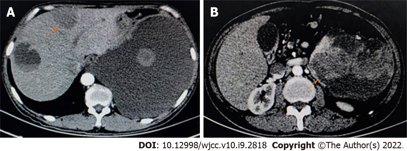
Figure 4 Plain and enhanced computed tomography revealed multiple round shadows of low density in the spleen and liver.
A: Computed tomography scan revealed that, in the left and right liver parenchyma, circular hypodensity reduction was observed with uneven density. On enhanced scan, the solid components showed slight enhancement, and no enhancement of hypodensity was observed. Massive pleural effusion; B: The spleen was enlarged, and multiple abnormal cystic solid density shadows were observed in and around the spleen. Enhanced scanning showed mild enhancement and partial fusion. The orange arrows indicate circular hypo-density regions.
- Citation: Pan D, Li TP, Xiong JH, Wang SB, Chen YX, Li JF, Xiao Q. Treatment with sorafenib plus camrelizumab after splenectomy for primary splenic angiosarcoma with liver metastasis: A case report and literature review. World J Clin Cases 2022; 10(9): 2818-2828
- URL: https://www.wjgnet.com/2307-8960/full/v10/i9/2818.htm
- DOI: https://dx.doi.org/10.12998/wjcc.v10.i9.2818


