Copyright
©The Author(s) 2022.
World J Clin Cases. Mar 26, 2022; 10(9): 2818-2828
Published online Mar 26, 2022. doi: 10.12998/wjcc.v10.i9.2818
Published online Mar 26, 2022. doi: 10.12998/wjcc.v10.i9.2818
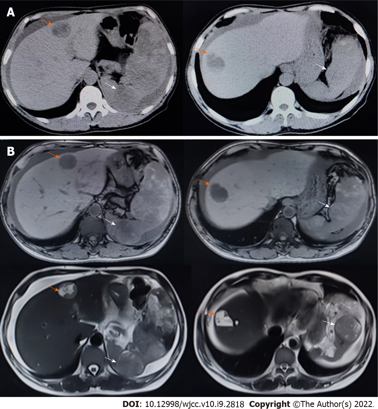
Figure 2 Computed tomography and magnetic resonance imaging performed soon after the first hospitalization.
Scale bar: 10 cm. A: We observed multiple masses in the liver and spleen on computed tomography; B: We observed multiple masses in the liver and spleen on magnetic resonance imaging. Orange arrows indicate masses in the liver, while the white arrows indicate masses in the spleen.
- Citation: Pan D, Li TP, Xiong JH, Wang SB, Chen YX, Li JF, Xiao Q. Treatment with sorafenib plus camrelizumab after splenectomy for primary splenic angiosarcoma with liver metastasis: A case report and literature review. World J Clin Cases 2022; 10(9): 2818-2828
- URL: https://www.wjgnet.com/2307-8960/full/v10/i9/2818.htm
- DOI: https://dx.doi.org/10.12998/wjcc.v10.i9.2818


