Copyright
©The Author(s) 2022.
World J Clin Cases. Mar 26, 2022; 10(9): 2710-2720
Published online Mar 26, 2022. doi: 10.12998/wjcc.v10.i9.2710
Published online Mar 26, 2022. doi: 10.12998/wjcc.v10.i9.2710
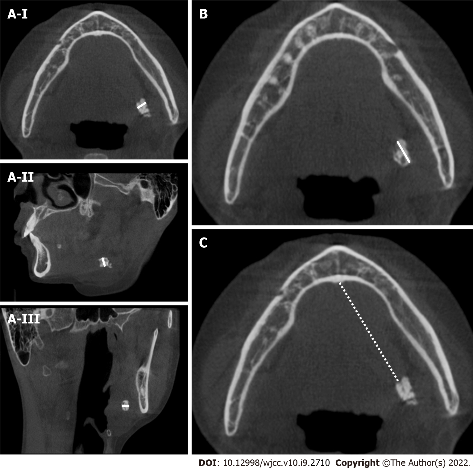
Figure 2 Measurements of the location, transverse diameter and longitudinal diameter of submandibular stones.
A-I: Measures the width of stones in the axial cone beam computed tomography (CBCT) views (white line); A-II: Measures the width of stones in the sagittal CBCT views (white line); A-III: Measures the width of stones in the coronal CBCT views (white line); B: Measures the longitudinal diameter of stones in the axial CBCT views (white line); C: Measures the distance between the anterior edge of the stone and the midpoint of the glossal cortex of the mandible in the axial CBCT views, which was defined as the location of the stone (white dotted line).
- Citation: Huang Y, Liang PS, Yang YC, Cai WX, Tao Q. Nomogram to predict the risk of endoscopic removal failure with forceps/baskets for treating submandibular stones. World J Clin Cases 2022; 10(9): 2710-2720
- URL: https://www.wjgnet.com/2307-8960/full/v10/i9/2710.htm
- DOI: https://dx.doi.org/10.12998/wjcc.v10.i9.2710


