Copyright
©The Author(s) 2022.
World J Clin Cases. Mar 16, 2022; 10(8): 2650-2656
Published online Mar 16, 2022. doi: 10.12998/wjcc.v10.i8.2650
Published online Mar 16, 2022. doi: 10.12998/wjcc.v10.i8.2650
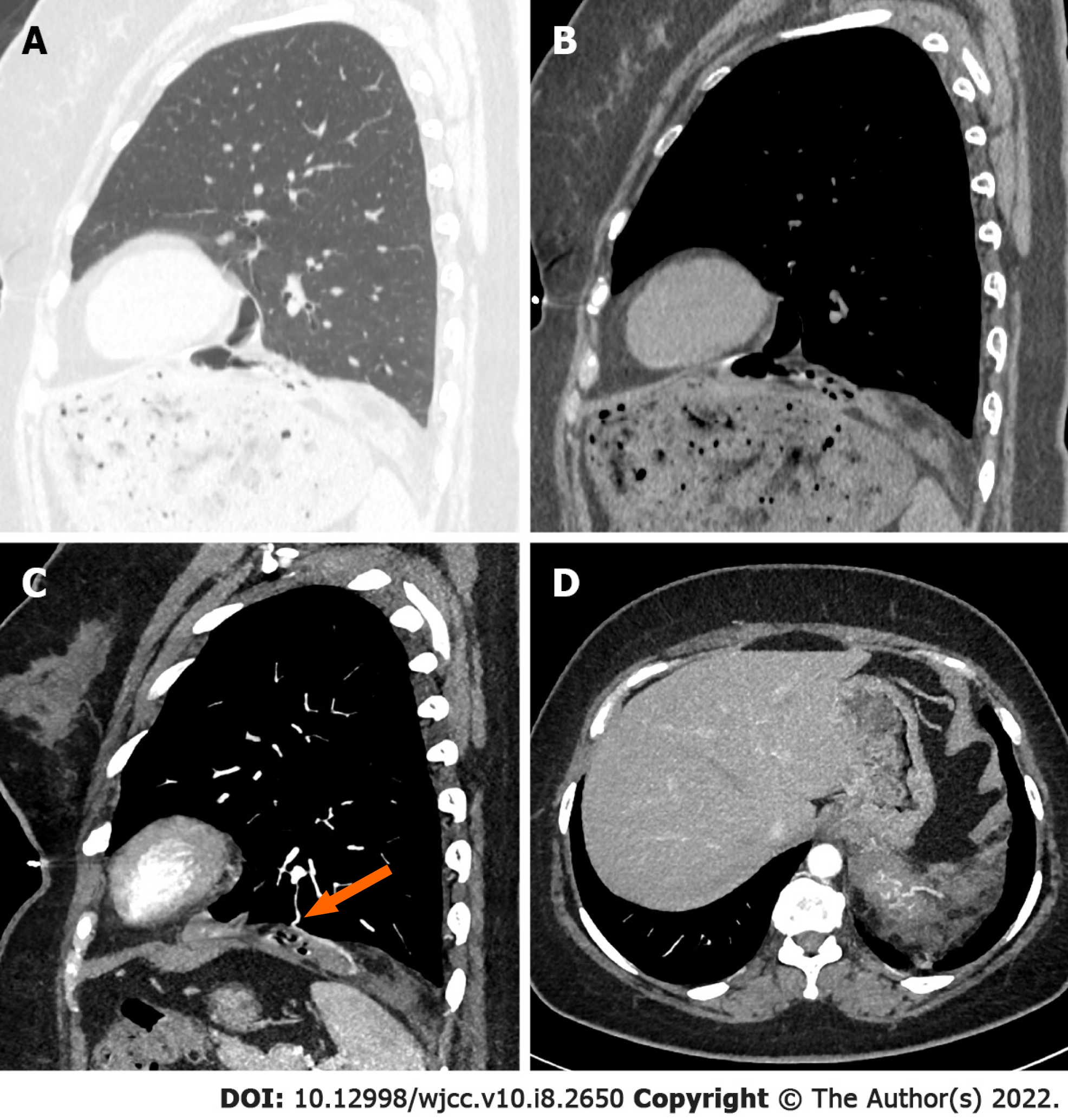
Figure 1 Computed tomography images of a large cystic-solid pulmonary hamartoma in a 53-year-old woman.
A and B: Nonenhanced chest computed tomography (CT) images show a well-defined tumor on the left diaphragm; C and D: Contrast-enhanced chest CT images show the blood supply of the tumor (solid arrow).
- Citation: Guo XW, Jia XD, Ji AD, Zhang DQ, Jia DZ, Zhang Q, Shao Q, Liu Y. Large cystic-solid pulmonary hamartoma: A case report. World J Clin Cases 2022; 10(8): 2650-2656
- URL: https://www.wjgnet.com/2307-8960/full/v10/i8/2650.htm
- DOI: https://dx.doi.org/10.12998/wjcc.v10.i8.2650


