Copyright
©The Author(s) 2022.
World J Clin Cases. Mar 16, 2022; 10(8): 2622-2628
Published online Mar 16, 2022. doi: 10.12998/wjcc.v10.i8.2622
Published online Mar 16, 2022. doi: 10.12998/wjcc.v10.i8.2622
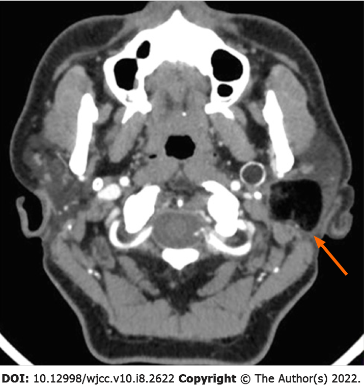
Figure 1 Axial-view contrast-enhanced computed tomography image.
The mass was located in the deep lobe of the left parotid gland. The medial part extended to the parapharyngeal space. Eggshell-like calcification was observed in the cyst wall. The cyst components were in different density, including a large amount of fat and a small number of keratinized substances.
- Citation: Liu HS, Zhang QY, Duan JF, Li G, Zhang J, Sun PF. Cystic teratoma of the parotid gland: A case report. World J Clin Cases 2022; 10(8): 2622-2628
- URL: https://www.wjgnet.com/2307-8960/full/v10/i8/2622.htm
- DOI: https://dx.doi.org/10.12998/wjcc.v10.i8.2622


