Copyright
©The Author(s) 2022.
World J Clin Cases. Mar 16, 2022; 10(8): 2610-2615
Published online Mar 16, 2022. doi: 10.12998/wjcc.v10.i8.2610
Published online Mar 16, 2022. doi: 10.12998/wjcc.v10.i8.2610
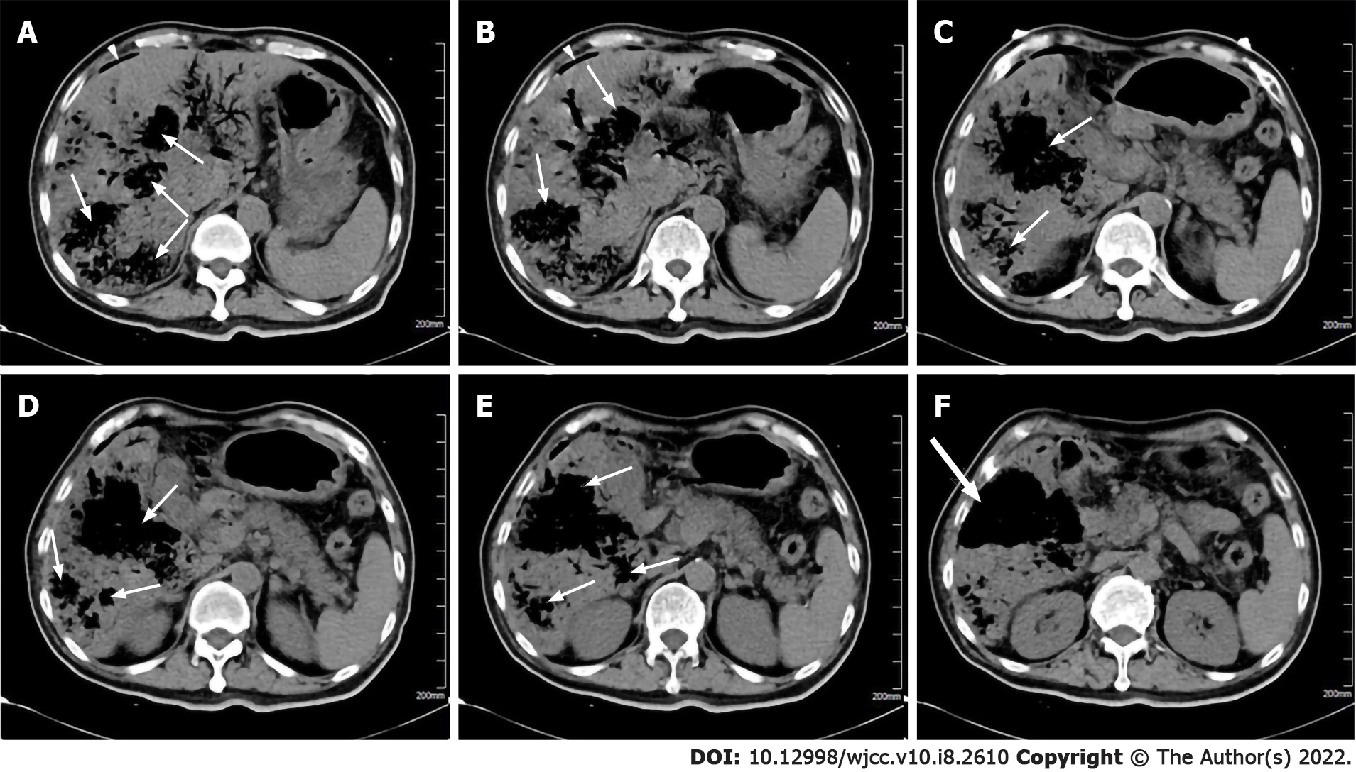
Figure 1 Abdominal computed tomography scan shows multiple emphysematous hepatic abscesses.
A and B: Arrowheads represent gas formation in the right subphrenic area; A-E: Thin arrows represent gas-containing liver abscess cavities; F: Thick arrows represent ruptured liver abscesses.
- Citation: Zhang JQ, He CC, Yuan B, Liu R, Qi YJ, Wang ZX, He XN, Li YM. Fatal systemic emphysematous infection caused by Klebsiella pneumoniae: A case report. World J Clin Cases 2022; 10(8): 2610-2615
- URL: https://www.wjgnet.com/2307-8960/full/v10/i8/2610.htm
- DOI: https://dx.doi.org/10.12998/wjcc.v10.i8.2610


