Copyright
©The Author(s) 2022.
World J Clin Cases. Mar 16, 2022; 10(8): 2516-2521
Published online Mar 16, 2022. doi: 10.12998/wjcc.v10.i8.2516
Published online Mar 16, 2022. doi: 10.12998/wjcc.v10.i8.2516
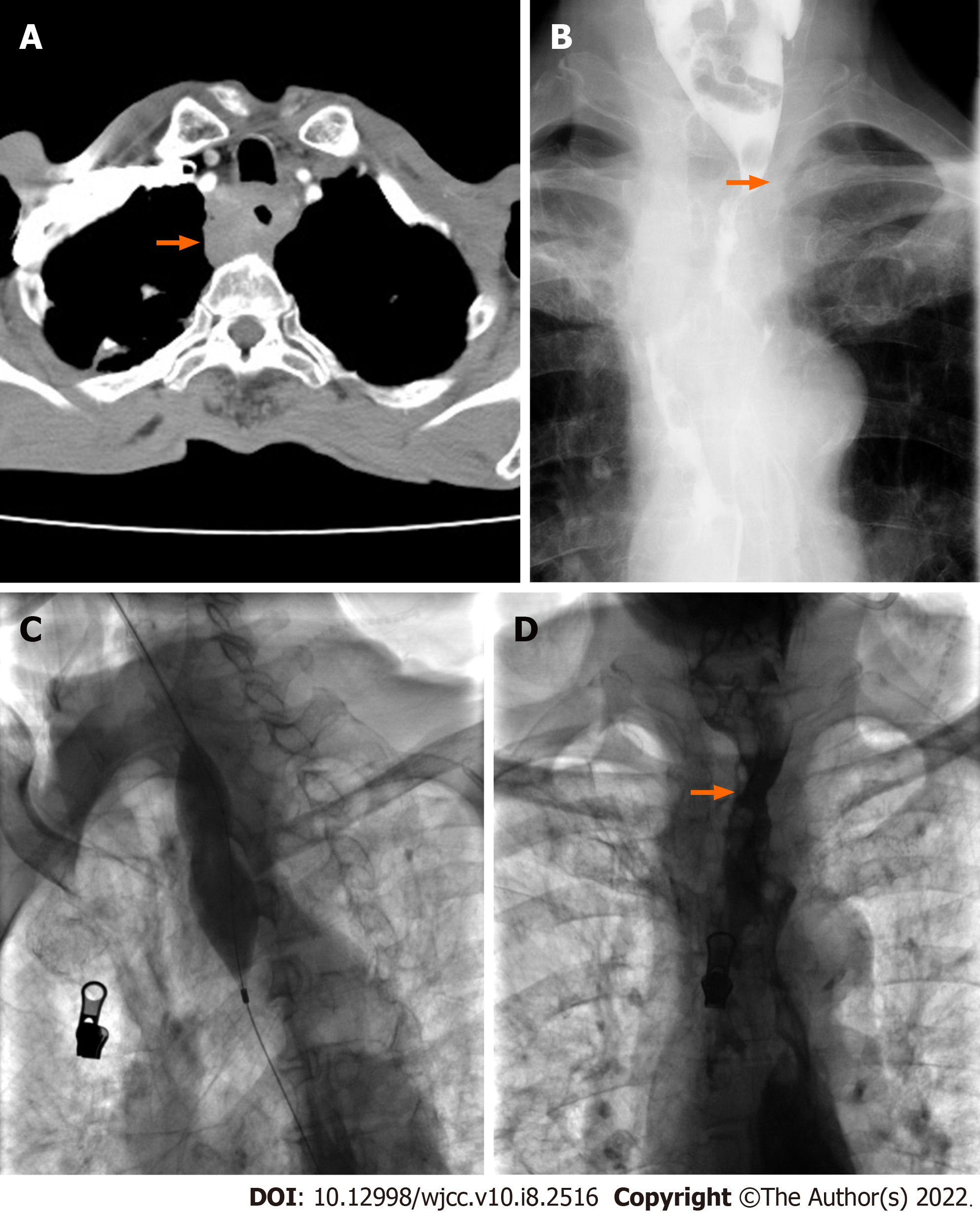
Figure 3 Balloon dilation performed approximately 40 d after stent removal.
A: Chest enhanced computed tomography examination showed obvious thickened esophageal wall (arrow); B: Esophagography showed severe stenosis (arrow) in the proximal part of the esophagus. C: A 28 mm in diameter and 40 mm in length big balloon catheter was used to dilate the stenotic section; D: Esophagography showed that the stenosis (arrow) disappeared after dilation.
- Citation: Bi YH, Ren JZ, Li JD, Han XW. Fluoroscopic removal of fractured, retained, embedded Z self-expanding metal stent using a guidewire lasso technique: A case report. World J Clin Cases 2022; 10(8): 2516-2521
- URL: https://www.wjgnet.com/2307-8960/full/v10/i8/2516.htm
- DOI: https://dx.doi.org/10.12998/wjcc.v10.i8.2516


