Copyright
©The Author(s) 2022.
World J Clin Cases. Mar 16, 2022; 10(8): 2516-2521
Published online Mar 16, 2022. doi: 10.12998/wjcc.v10.i8.2516
Published online Mar 16, 2022. doi: 10.12998/wjcc.v10.i8.2516
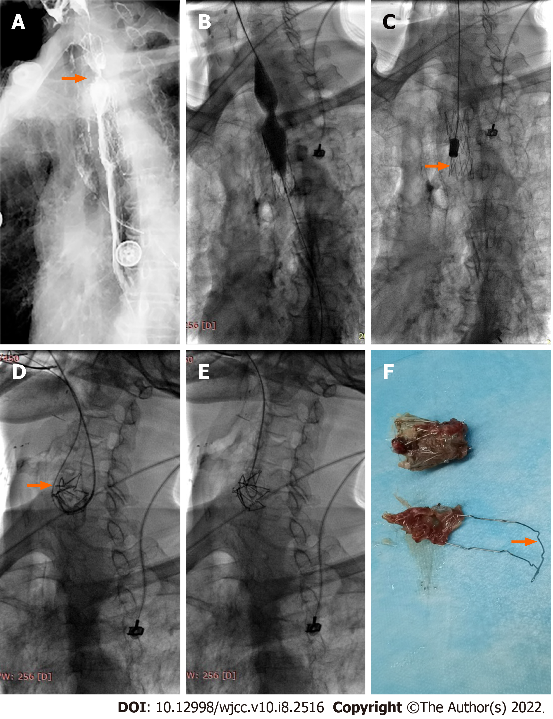
Figure 2 Balloon dilation and stent removal performed 13.
5 mo after stenting. A: Esophagography showed severe stenosis (arrow) above the proximal stent; B: A 20 mm in diameter and 40 mm in length balloon catheter was introduced to dilate the stenotic segment; C: A stent removal sheath was introduced and then a removal hook (arrow) was placed in the lower part of the stent, stent fracture occurred and only one-third of the stent was retrieved; D: A 0.035-inch guidewire was introduced and passed over the remaining fractured stent and looped back from the stent to the mouth (arrow); E: A suction tube was introduced through the guidewire and then the guidewire was grabbed, acting like a “lasso” on tightening; F: The fractured stent was successfully removed and the strut was deformed (arrow).
- Citation: Bi YH, Ren JZ, Li JD, Han XW. Fluoroscopic removal of fractured, retained, embedded Z self-expanding metal stent using a guidewire lasso technique: A case report. World J Clin Cases 2022; 10(8): 2516-2521
- URL: https://www.wjgnet.com/2307-8960/full/v10/i8/2516.htm
- DOI: https://dx.doi.org/10.12998/wjcc.v10.i8.2516


