Copyright
©The Author(s) 2022.
World J Clin Cases. Mar 16, 2022; 10(8): 2516-2521
Published online Mar 16, 2022. doi: 10.12998/wjcc.v10.i8.2516
Published online Mar 16, 2022. doi: 10.12998/wjcc.v10.i8.2516
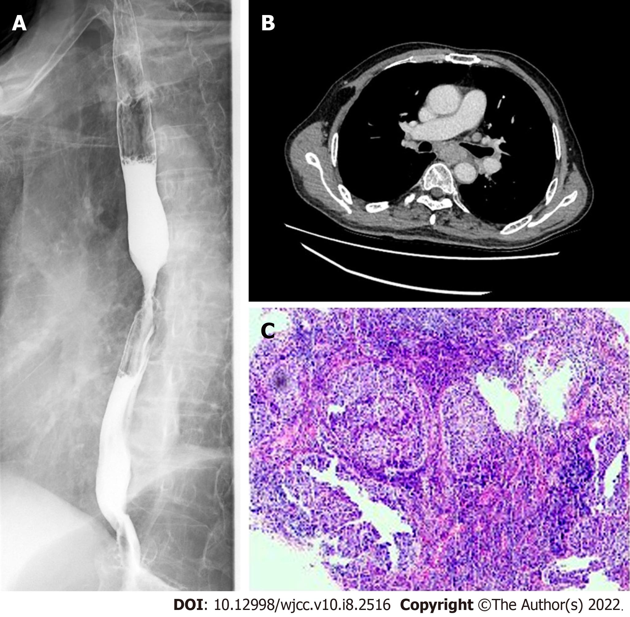
Figure 1 Examination before esophagectomy 21 mo ago.
A: Esophagography showed that the tumor was located in the middle and lower part of the esophagus; B: A chest contrast-enhanced computed tomography scan showed thickened esophageal wall; C: Esophageal gastroscopy examination was performed and biopsy pathology confirmed esophageal squamous cell carcinoma.
- Citation: Bi YH, Ren JZ, Li JD, Han XW. Fluoroscopic removal of fractured, retained, embedded Z self-expanding metal stent using a guidewire lasso technique: A case report. World J Clin Cases 2022; 10(8): 2516-2521
- URL: https://www.wjgnet.com/2307-8960/full/v10/i8/2516.htm
- DOI: https://dx.doi.org/10.12998/wjcc.v10.i8.2516


