Copyright
©The Author(s) 2022.
World J Clin Cases. Mar 16, 2022; 10(8): 2510-2515
Published online Mar 16, 2022. doi: 10.12998/wjcc.v10.i8.2510
Published online Mar 16, 2022. doi: 10.12998/wjcc.v10.i8.2510
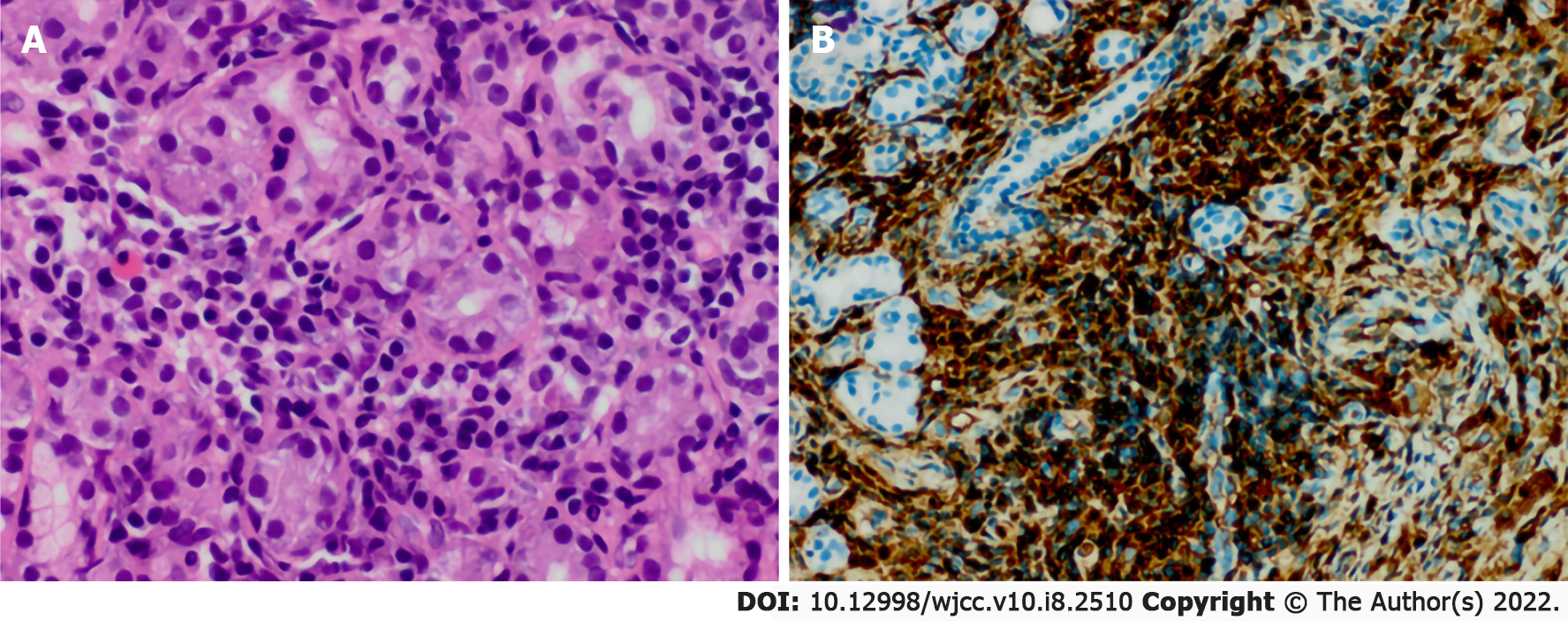
Figure 2 Histopathology of the lymph node.
A: The section shows dense lymphoplasmacytic cells with fibrotic changes (H&E 400×); B: Immunohistochemical staining shows an average of 100 IgG4-positive plasma cells per high-power field.
- Citation: He JW, Zou QM, Pan J, Wang SS, Xiang ST. Immunoglobulin G4-related kidney disease involving the renal pelvis and perirenal fat: A case report. World J Clin Cases 2022; 10(8): 2510-2515
- URL: https://www.wjgnet.com/2307-8960/full/v10/i8/2510.htm
- DOI: https://dx.doi.org/10.12998/wjcc.v10.i8.2510


