Copyright
©The Author(s) 2022.
World J Clin Cases. Mar 16, 2022; 10(8): 2504-2509
Published online Mar 16, 2022. doi: 10.12998/wjcc.v10.i8.2504
Published online Mar 16, 2022. doi: 10.12998/wjcc.v10.i8.2504
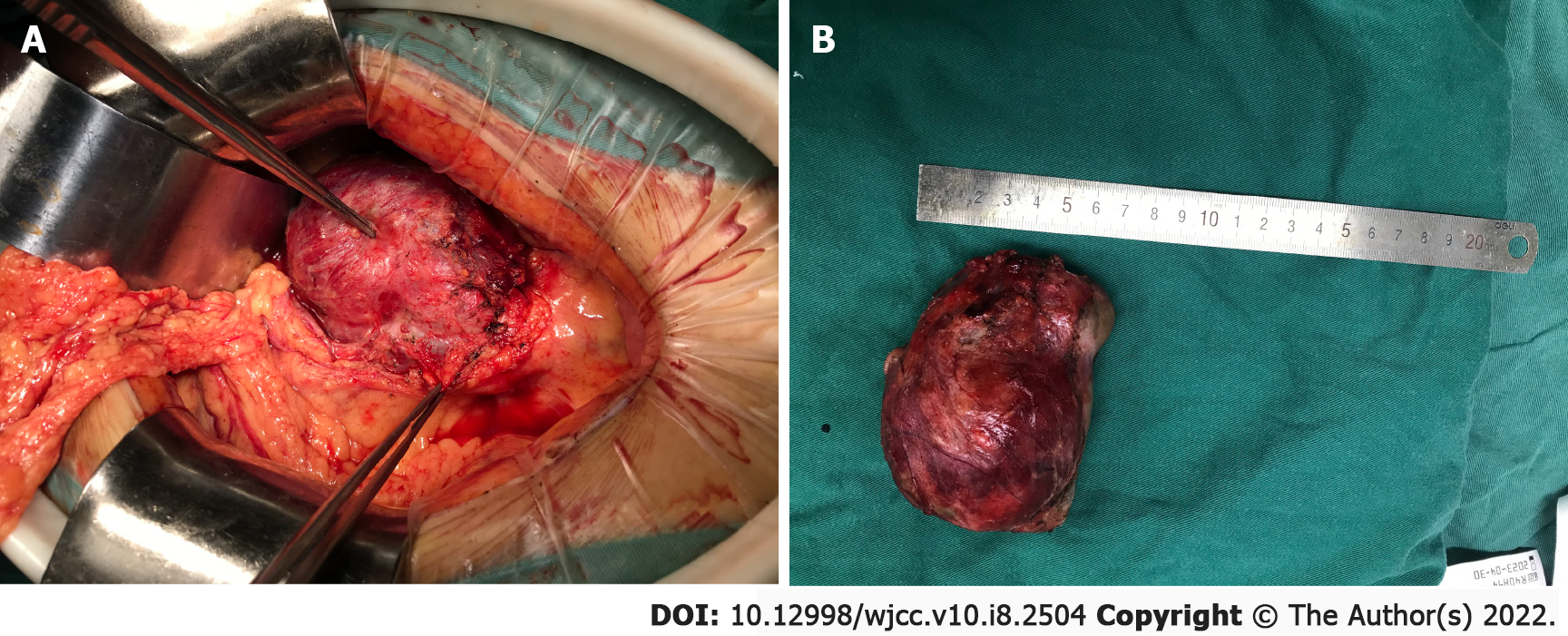
Figure 2 Intraoperative view of gross inspection of the cystic mass.
A: Large retroperitoneal mass is closely connected to the superior mesenteric vein and to the root of the superior mesenteric artery; B: Operative specimen showing a giant retroperitoneal cystic mass of approximately 10 cm in the largest dimension.
- Citation: Ma J, Zhang YM, Zhou CP, Zhu L. Retroperitoneal congenital epidermoid cyst misdiagnosed as a solid pseudopapillary tumor of the pancreas: A case report. World J Clin Cases 2022; 10(8): 2504-2509
- URL: https://www.wjgnet.com/2307-8960/full/v10/i8/2504.htm
- DOI: https://dx.doi.org/10.12998/wjcc.v10.i8.2504


