Copyright
©The Author(s) 2022.
World J Clin Cases. Mar 16, 2022; 10(8): 2484-2490
Published online Mar 16, 2022. doi: 10.12998/wjcc.v10.i8.2484
Published online Mar 16, 2022. doi: 10.12998/wjcc.v10.i8.2484
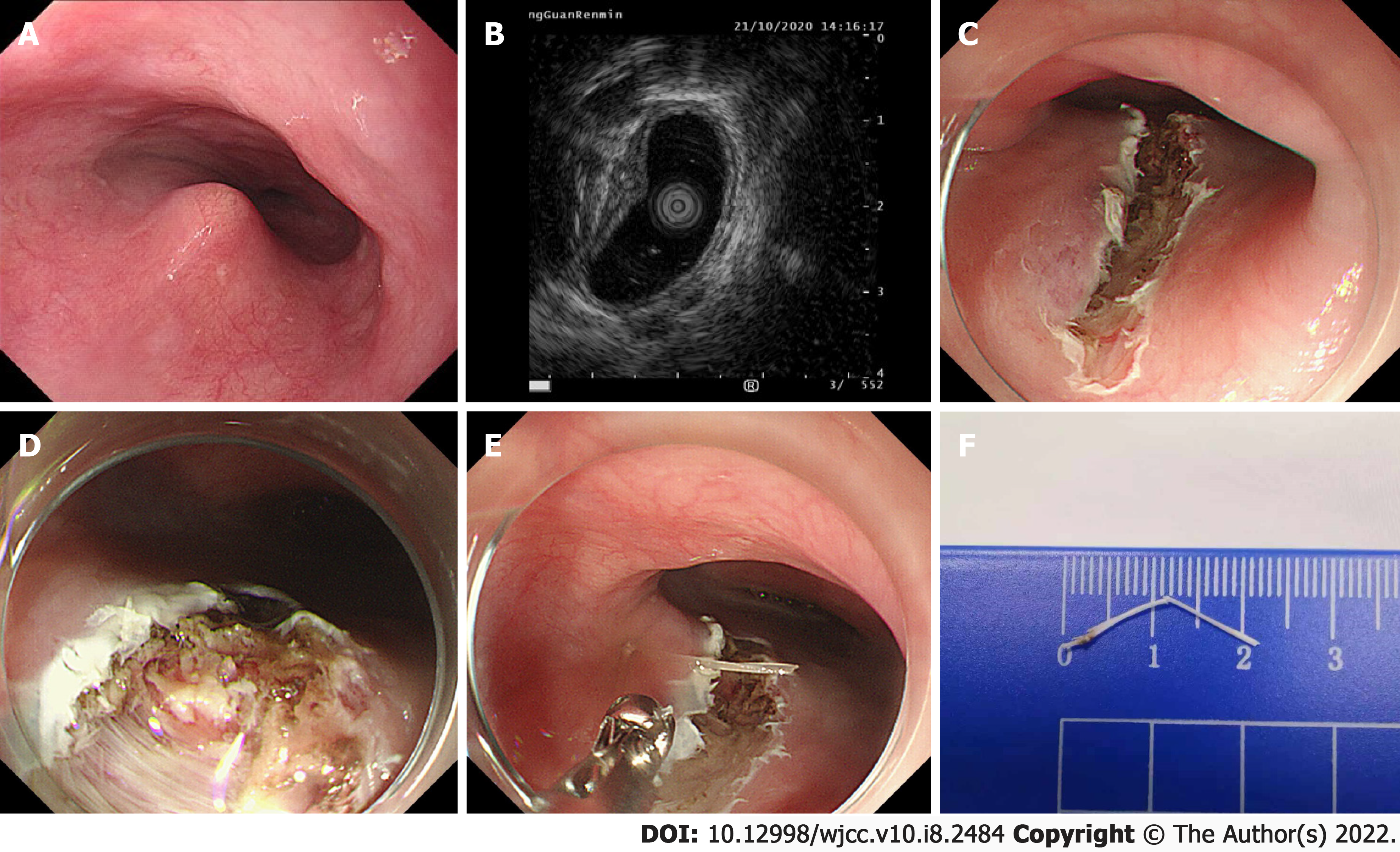
Figure 2 Images of endoscopy operation.
A: Endoscopic examination was performed on October 21, 2020, and a bulge was identified; B: Endoscopic ultrasonography showed a strong echo light cluster protruding beyond the muscularis propria under the bulge; C: The nodule was incised with a dual knife and IT knife to the deep part of the esophageal muscularis; D: A suspicious fishbone with a length of about 1.5 mm was found on the distal side of the nodule; E: The fishbone was pulled out with biopsy forceps; F: The total length of the fishbone was approximately 22 mm.
- Citation: Chen ZC, Chen GQ, Chen XC, Zheng CY, Cao WD, Deng GH. Endoscopic extraction of a submucosal esophageal foreign body piercing into the thoracic aorta: A case report. World J Clin Cases 2022; 10(8): 2484-2490
- URL: https://www.wjgnet.com/2307-8960/full/v10/i8/2484.htm
- DOI: https://dx.doi.org/10.12998/wjcc.v10.i8.2484


