Copyright
©The Author(s) 2022.
World J Clin Cases. Mar 16, 2022; 10(8): 2484-2490
Published online Mar 16, 2022. doi: 10.12998/wjcc.v10.i8.2484
Published online Mar 16, 2022. doi: 10.12998/wjcc.v10.i8.2484
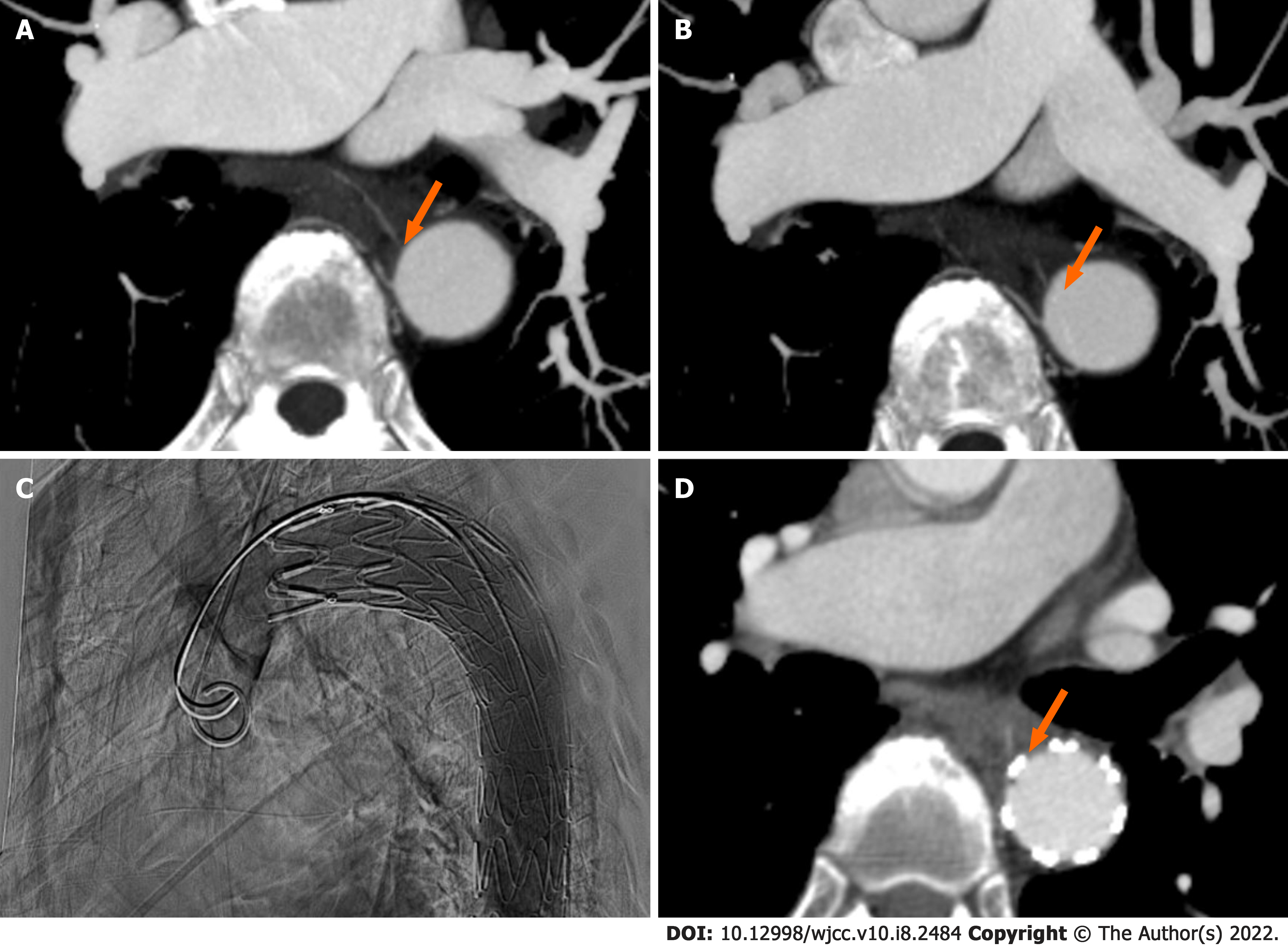
Figure 1 Computed tomography images and thoracic endovascular aortic repair images.
A: Emergency thoracic computed tomography (CT) angiography revealed a fishbone in the esophagus involving the wall of the thoracic aorta; B: Enhanced CT angiography revealed that the fishbone had directly penetrated the thoracic aorta after esophagoscopic examination; C: On October 18, 2020, we successfully performed thoracic endovascular aortic repair (TEVAR); D: CT angiography after TEVAR showed that the fishbone was pressed against the edge of the blood vessel.
- Citation: Chen ZC, Chen GQ, Chen XC, Zheng CY, Cao WD, Deng GH. Endoscopic extraction of a submucosal esophageal foreign body piercing into the thoracic aorta: A case report. World J Clin Cases 2022; 10(8): 2484-2490
- URL: https://www.wjgnet.com/2307-8960/full/v10/i8/2484.htm
- DOI: https://dx.doi.org/10.12998/wjcc.v10.i8.2484


