Copyright
©The Author(s) 2022.
World J Clin Cases. Mar 16, 2022; 10(8): 2404-2419
Published online Mar 16, 2022. doi: 10.12998/wjcc.v10.i8.2404
Published online Mar 16, 2022. doi: 10.12998/wjcc.v10.i8.2404
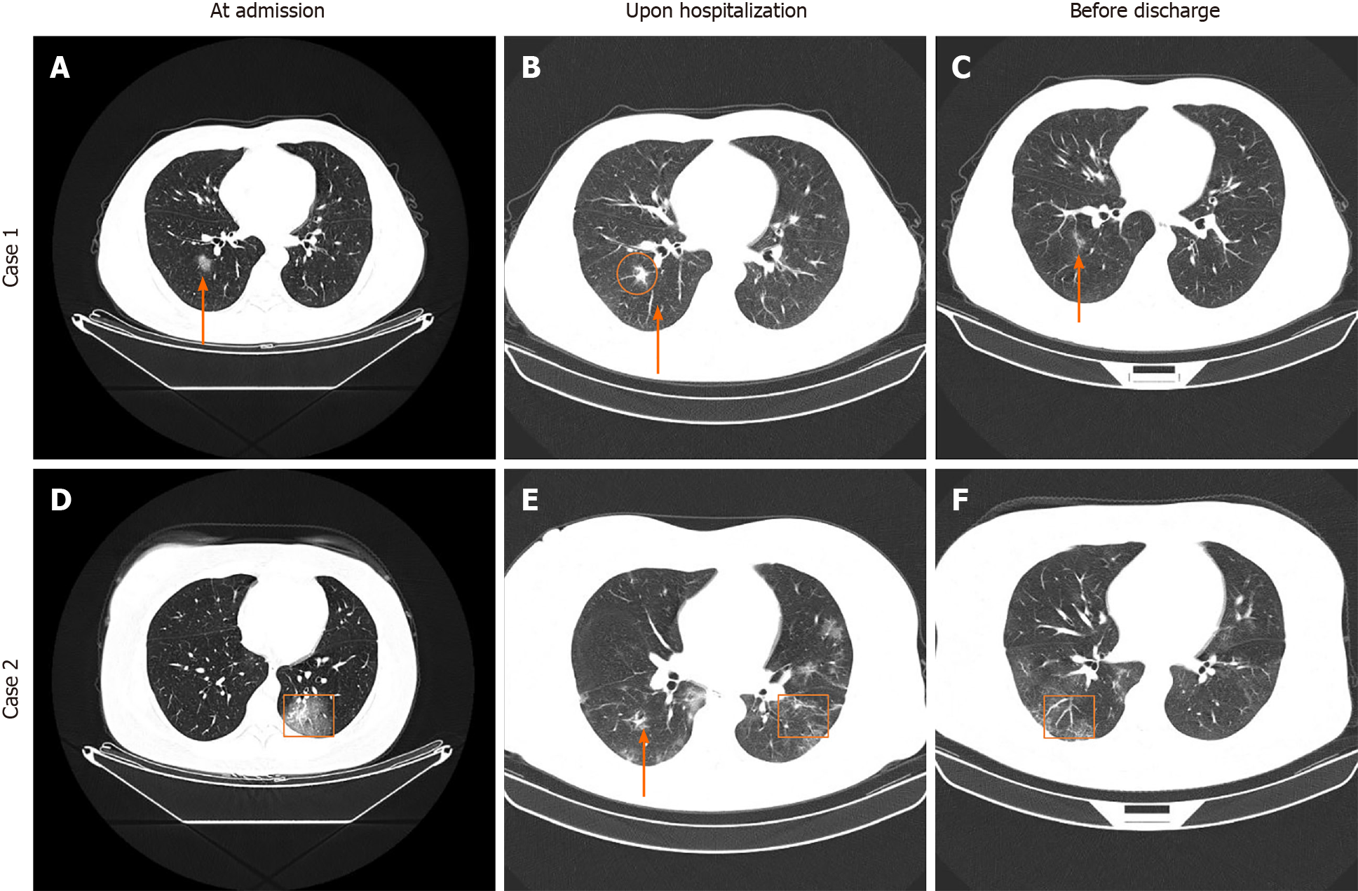
Figure 9 Representative computed tomography images of two patients with coronavirus disease 2019 at admission, upon hospitalization and before discharge.
A: Punctate ground-glass opacity (GGO) was observed in the inferior lobe of the right lung; B: Partial absorption was observed for the GGO lesion in the inferior lobe of the right lung; C: A striped high-density shade was only observed in the inferior lobe of the right lung after further absorption; D: Patchy GGO was observed in the inferior lobe of the left lung; E: Multiple patchy GGOs were observed in both lungs, and the number of lesions was increased; F: Remarkable absorption of patchy GGOs was observed in both lungs. The red boxes and arrows indicate abnormalities.
- Citation: Nie XB, Shi BS, Zhang L, Niu WL, Xue T, Li LQ, Wei XY, Wang YD, Chen WD, Hou RF. Epidemiological features and dynamic changes in blood biochemical indices for COVID-19 patients in Hebi. World J Clin Cases 2022; 10(8): 2404-2419
- URL: https://www.wjgnet.com/2307-8960/full/v10/i8/2404.htm
- DOI: https://dx.doi.org/10.12998/wjcc.v10.i8.2404


