Copyright
©The Author(s) 2022.
World J Clin Cases. Mar 6, 2022; 10(7): 2341-2350
Published online Mar 6, 2022. doi: 10.12998/wjcc.v10.i7.2341
Published online Mar 6, 2022. doi: 10.12998/wjcc.v10.i7.2341
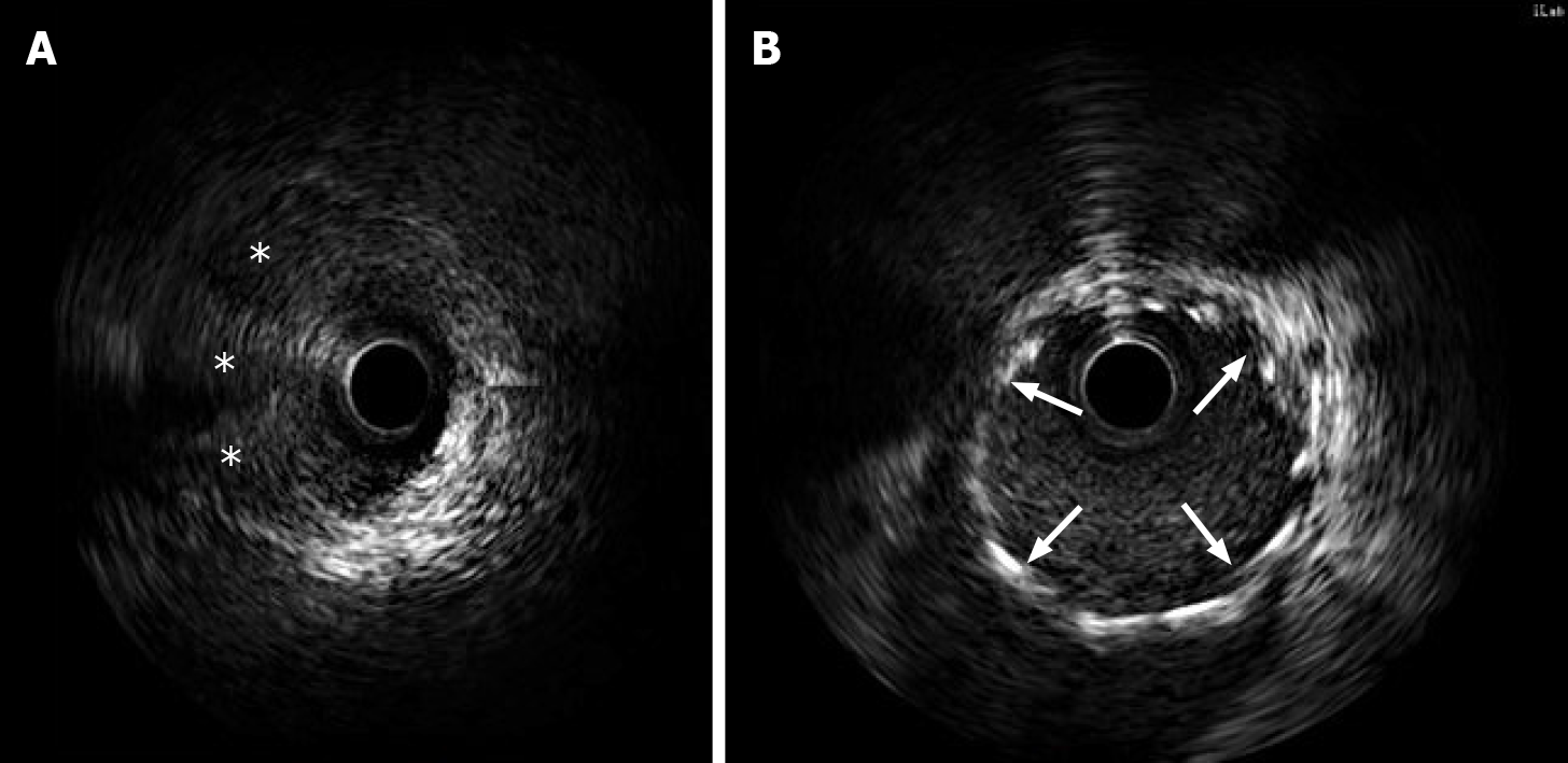
Figure 3 Intravascular ultrasound confirmed a false lumen with a prominent dissection from the left main trunk coronary artery to the ostium of the left anterior descending artery.
A: Intravascular ultrasound (IVUS) images showed artery dissection starting from the left main trunk coronary artery to the ostium of the left anterior descending artery. The guidewire passed through the true lumen properly; B: IVUS demonstrated stents attached to the endothelium well that covered the artery dissections completely. Intramural hematoma (stellate). The implanted stents are indicated with arrows.
- Citation: Liu SF, Zhao YN, Jia CW, Ma TY, Cai SD, Gao F. Spontaneous dissection of proximal left main coronary artery in a healthy adolescent presenting with syncope: A case report. World J Clin Cases 2022; 10(7): 2341-2350
- URL: https://www.wjgnet.com/2307-8960/full/v10/i7/2341.htm
- DOI: https://dx.doi.org/10.12998/wjcc.v10.i7.2341


