Copyright
©The Author(s) 2022.
World J Clin Cases. Mar 6, 2022; 10(7): 2330-2335
Published online Mar 6, 2022. doi: 10.12998/wjcc.v10.i7.2330
Published online Mar 6, 2022. doi: 10.12998/wjcc.v10.i7.2330
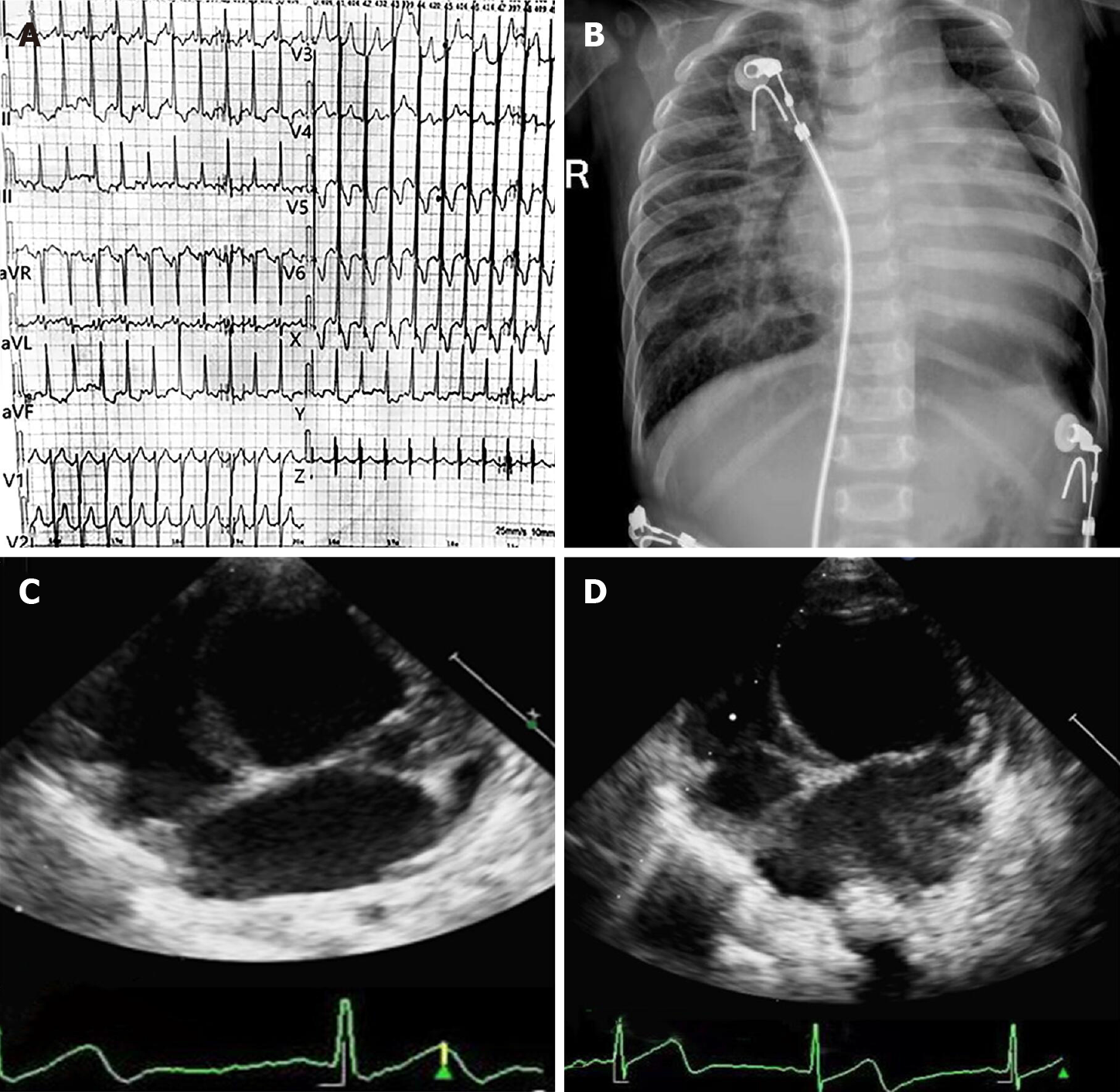
Figure 1 Electrocardiography and imaging examinations of the patient.
A: Abnormal 12-lead electrocardiography indicated high voltages in the left precordial leads and diffuse T wave inversion; B: Chest radiography demonstrated cardiac enlargement and pulmonary congestion; C: Dilated left ventricle approximately 43 mm in late diastole and reduced ejection fraction approximately 26% (echocardiography at admission); D: Dilated left ventricle approximately 46 mm in late diastole, reduced ejection fraction approximately 27% (echocardiography at a follow-up of 6 mo).
- Citation: Jiang P, Xiao L, Guo Y, Hu R, Zhang BY, He Y. Novel mutations of the Alström syndrome 1 gene in an infant with dilated cardiomyopathy: A case report. World J Clin Cases 2022; 10(7): 2330-2335
- URL: https://www.wjgnet.com/2307-8960/full/v10/i7/2330.htm
- DOI: https://dx.doi.org/10.12998/wjcc.v10.i7.2330


