Copyright
©The Author(s) 2022.
World J Clin Cases. Mar 6, 2022; 10(7): 2322-2329
Published online Mar 6, 2022. doi: 10.12998/wjcc.v10.i7.2322
Published online Mar 6, 2022. doi: 10.12998/wjcc.v10.i7.2322
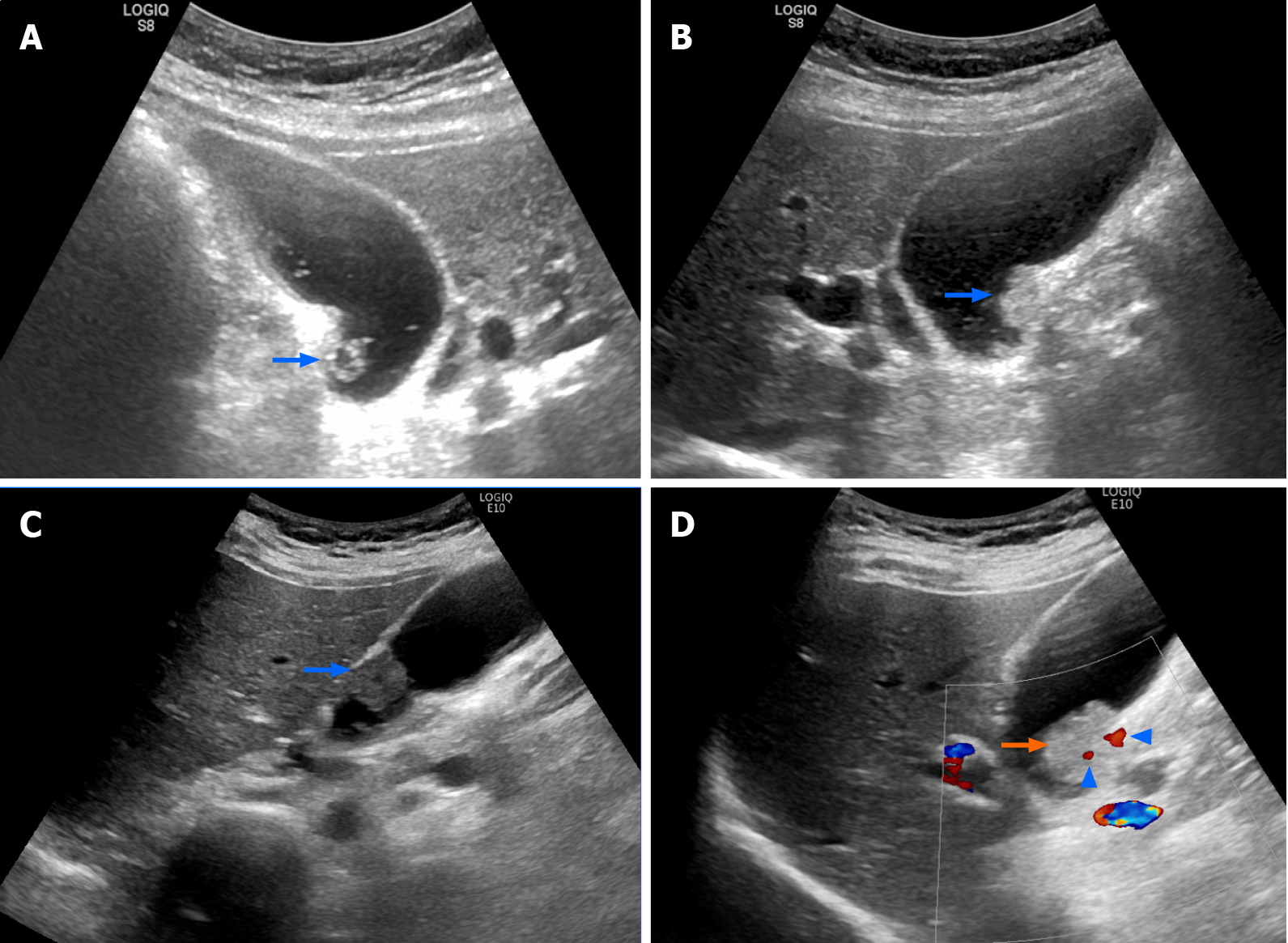
Figure 2 Evolutionary change in gall bladder polyp.
A: A 1-cm hyperechoic lesion with hypoechoic component (polyp) at the gall bladder wall (arrow); B: The polyp became larger compared to its size 1 year prior (arrow); C: The polyp became hypoechoic (arrow) after another 6 mo; D: Ultrasound performed just before surgery revealed a Doppler signal (arrowhead) within the tumor (arrow).
- Citation: Hsiao TH, Wu CC, Tseng HH, Chen JH. Synchronous but separate neuroendocrine tumor and high-grade dysplasia/adenoma of the gall bladder: A case report. World J Clin Cases 2022; 10(7): 2322-2329
- URL: https://www.wjgnet.com/2307-8960/full/v10/i7/2322.htm
- DOI: https://dx.doi.org/10.12998/wjcc.v10.i7.2322


