Copyright
©The Author(s) 2022.
World J Clin Cases. Mar 6, 2022; 10(7): 2275-2280
Published online Mar 6, 2022. doi: 10.12998/wjcc.v10.i7.2275
Published online Mar 6, 2022. doi: 10.12998/wjcc.v10.i7.2275
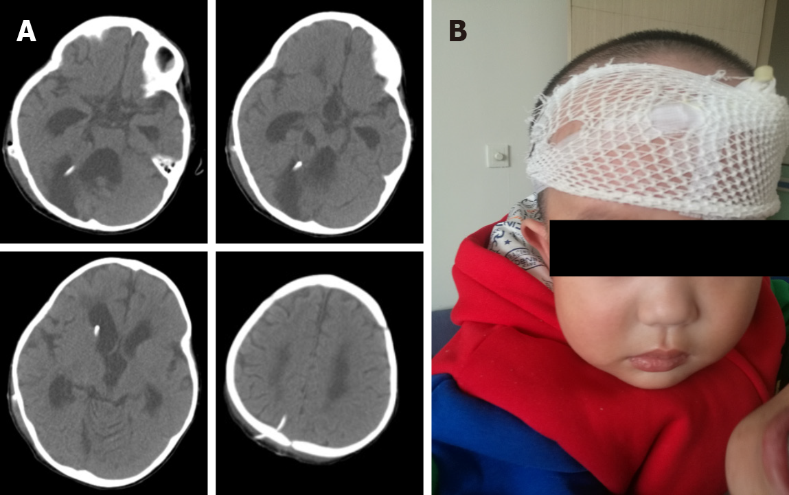
Figure 3 Postoperative imaging examination and child status.
A: Postoperative brain computed tomography showing shrunk lateral ventricles and disappeared capped edema at the bilateral frontal angle; the head ends of the drainage tube in the lateral ventricle and posterior fossa cysts were well-positioned; B: Postoperative status of the child: Brain computed tomography at 1 mo postoperatively revealed significantly shrunk lateral ventricle; the head ends of the drainage tube in the lateral ventricle and posterior fossa cysts were well-positioned. The cysts were significantly shrunk and there was cerebellar vermis dysplasia.
- Citation: Dong ZQ, Jia YF, Gao ZS, Li Q, Niu L, Yang Q, Pan YW, Li Q. Y-shaped shunt for the treatment of Dandy-Walker malformation combined with giant arachnoid cysts: A case report. World J Clin Cases 2022; 10(7): 2275-2280
- URL: https://www.wjgnet.com/2307-8960/full/v10/i7/2275.htm
- DOI: https://dx.doi.org/10.12998/wjcc.v10.i7.2275


