Copyright
©The Author(s) 2022.
World J Clin Cases. Mar 6, 2022; 10(7): 2229-2246
Published online Mar 6, 2022. doi: 10.12998/wjcc.v10.i7.2229
Published online Mar 6, 2022. doi: 10.12998/wjcc.v10.i7.2229
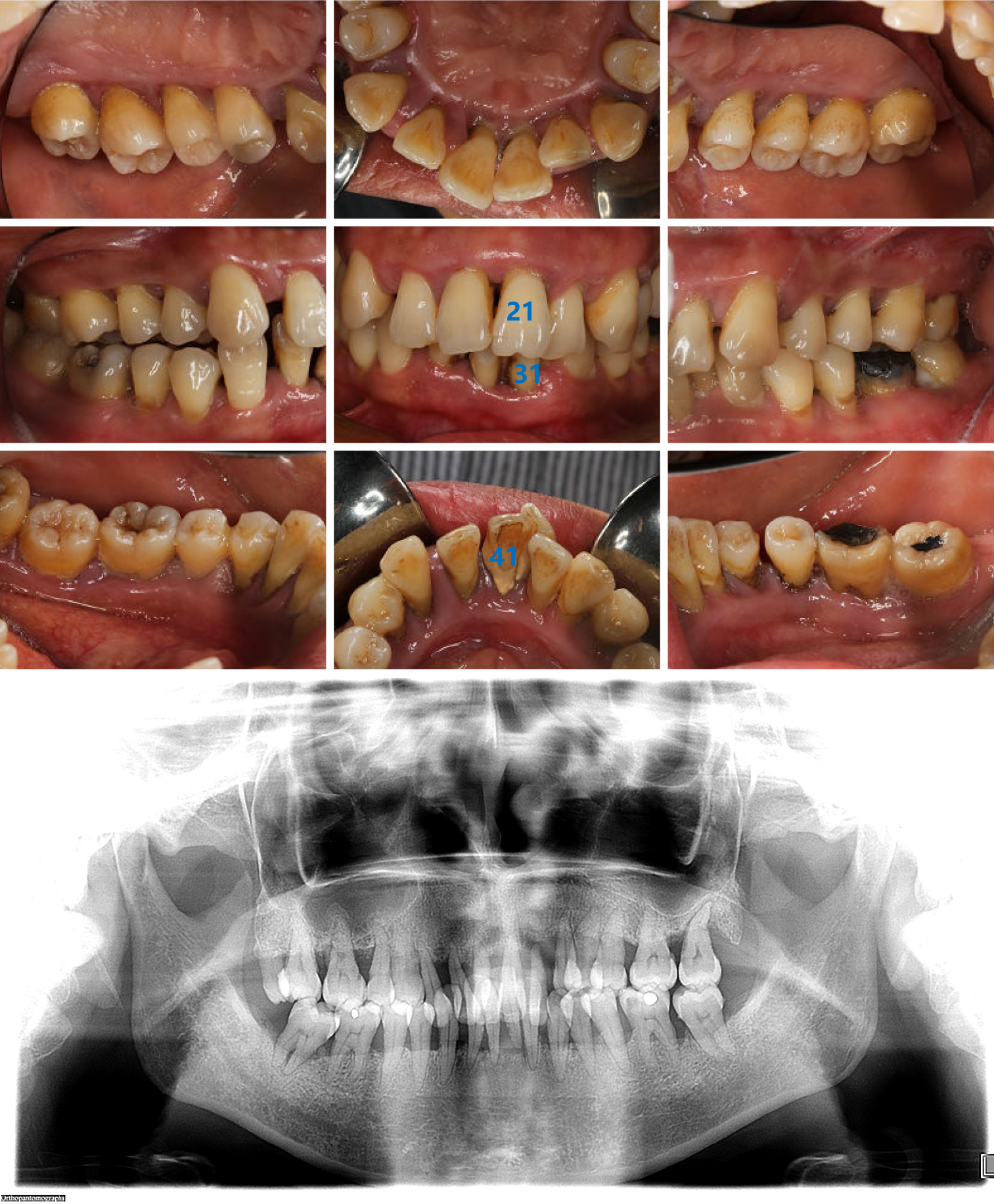
Figure 1 Intraoral images and radiographic image obtained at the first visit.
Intraoral images show the poor level of oral hygiene, a large amount of dental calculus, and obvious plaque retention. No. 21 tooth was distinctly labially inclined and Nos. 31 and 41 were extremely loose. The radiographic image reveals that the full mouth alveolar bone was absorbed to varying degrees into the middle 1/2 and apical 1/3 of the root, with Nos. 22 and 31 absorbed to the apical region.
- Citation: Li LJ, Yan X, Yu Q, Yan FH, Tan BC. Multidisciplinary non-surgical treatment of advanced periodontitis: A case report. World J Clin Cases 2022; 10(7): 2229-2246
- URL: https://www.wjgnet.com/2307-8960/full/v10/i7/2229.htm
- DOI: https://dx.doi.org/10.12998/wjcc.v10.i7.2229


