Copyright
©The Author(s) 2022.
World J Clin Cases. Mar 6, 2022; 10(7): 2087-2094
Published online Mar 6, 2022. doi: 10.12998/wjcc.v10.i7.2087
Published online Mar 6, 2022. doi: 10.12998/wjcc.v10.i7.2087
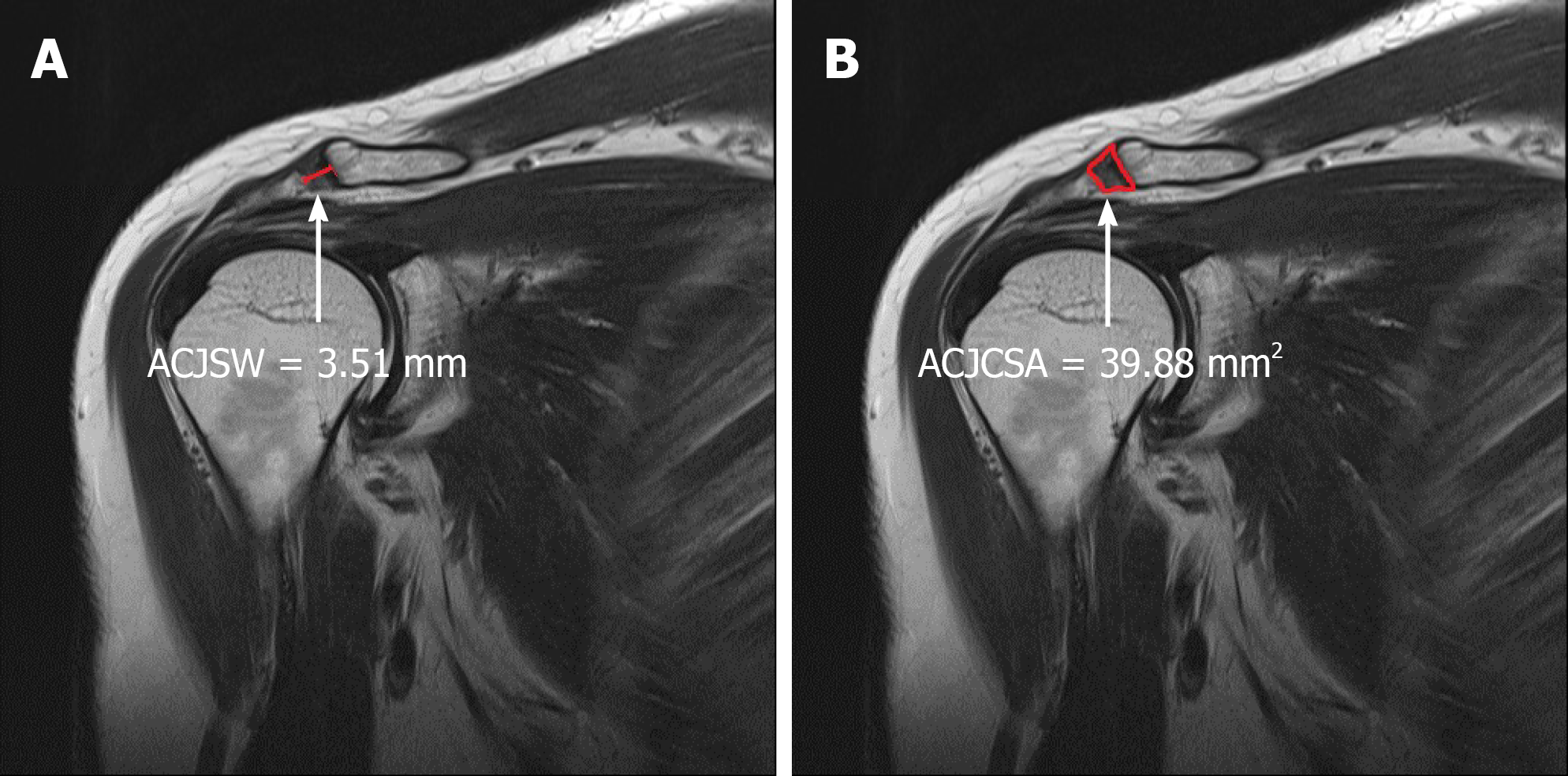
Figure 1 Measurement of both the acromioclavicular joint space width (A) and acromioclavicular joint cross-sectional area (B) in the normal control group was carried out on coronal T2-weighted shoulder-MR acromioclavicular joint images.
- Citation: Joo Y, Moon JY, Han JY, Bang YS, Kang KN, Lim YS, Choi YS, Kim YU. Usefulness of the acromioclavicular joint cross-sectional area as a diagnostic image parameter of acromioclavicular osteoarthritis. World J Clin Cases 2022; 10(7): 2087-2094
- URL: https://www.wjgnet.com/2307-8960/full/v10/i7/2087.htm
- DOI: https://dx.doi.org/10.12998/wjcc.v10.i7.2087


