Copyright
©The Author(s) 2022.
World J Clin Cases. Feb 26, 2022; 10(6): 2036-2044
Published online Feb 26, 2022. doi: 10.12998/wjcc.v10.i6.2036
Published online Feb 26, 2022. doi: 10.12998/wjcc.v10.i6.2036
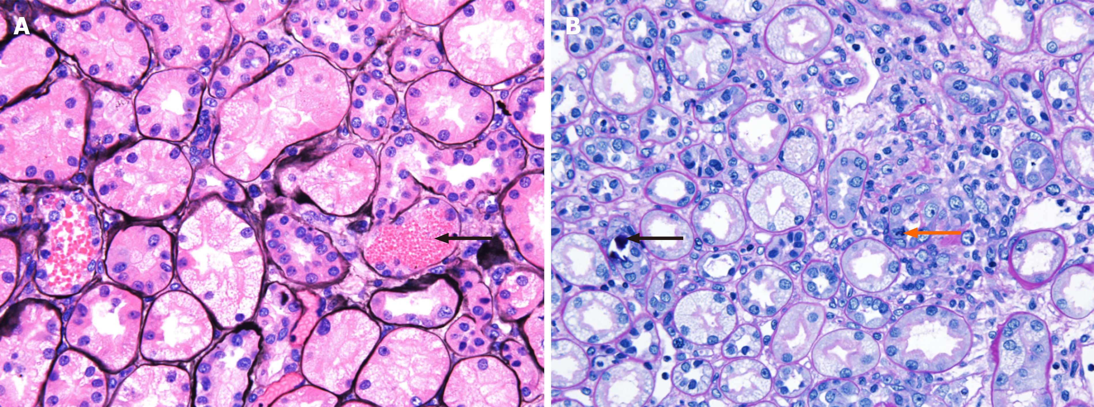
Figure 2 Light micrographs of renal biopsy.
A: The tubules show vacuolated degeneration with some red blood cells, granular materials (black arrow) (methenamine silver stain, × 400); B: The tubules show calcium concretions (black arrow) in tubular lumina and mitosis (orange arrow) (periodic acid-Schiff stain, × 400).
- Citation: Park S, Ryu HS, Lee JK, Park SS, Kwon SJ, Hwang WM, Yun SR, Park MH, Park Y. Acute kidney injury due to intravenous detergent poisoning: A case report. World J Clin Cases 2022; 10(6): 2036-2044
- URL: https://www.wjgnet.com/2307-8960/full/v10/i6/2036.htm
- DOI: https://dx.doi.org/10.12998/wjcc.v10.i6.2036


