Copyright
©The Author(s) 2022.
World J Clin Cases. Feb 26, 2022; 10(6): 2023-2029
Published online Feb 26, 2022. doi: 10.12998/wjcc.v10.i6.2023
Published online Feb 26, 2022. doi: 10.12998/wjcc.v10.i6.2023
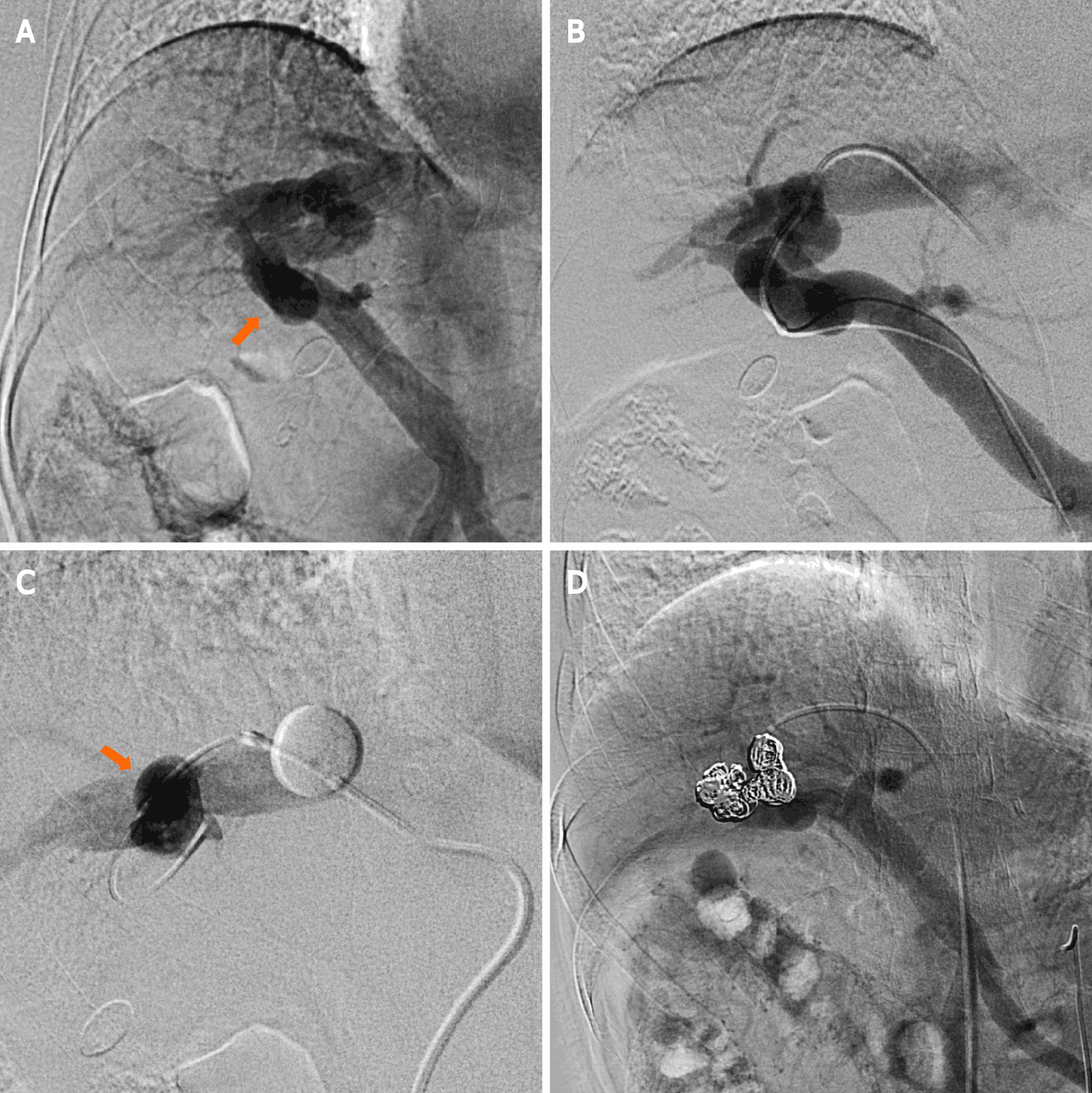
Figure 2 Superior mesenteric arterial portography image.
A: Superior mesenteric arterial portography image shows communication between the right portal and hepatic veins (arrow); B: Connections between the right portal and hepatic veins are observed following the injection of contrast media via a micro-catheter that was advanced into the portal vein; C: The right hepatic vein is occluded by the balloon catheter to decrease the hepatofugal blood flow into the intrahepatic portosystemic shunt (IPSVS), and the IPSVS is observed with balloon-occluded retrograde right hepatic venography (arrow); D: Superior mesenteric arterial portography after the procedure shows that the IPSVS is sufficiently occupied by coils, and there is no residual hepatofugal blood flow into the IPSVS.
- Citation: Saito H, Murata S, Sugihara F, Ueda T, Yasui D, Miki I, Hayashi H, Kumita SI. Successful embolization of an intrahepatic portosystemic shunt using balloon-occluded retrograde transvenous obliteration: A case report. World J Clin Cases 2022; 10(6): 2023-2029
- URL: https://www.wjgnet.com/2307-8960/full/v10/i6/2023.htm
- DOI: https://dx.doi.org/10.12998/wjcc.v10.i6.2023


