Copyright
©The Author(s) 2022.
World J Clin Cases. Feb 26, 2022; 10(6): 1981-1990
Published online Feb 26, 2022. doi: 10.12998/wjcc.v10.i6.1981
Published online Feb 26, 2022. doi: 10.12998/wjcc.v10.i6.1981
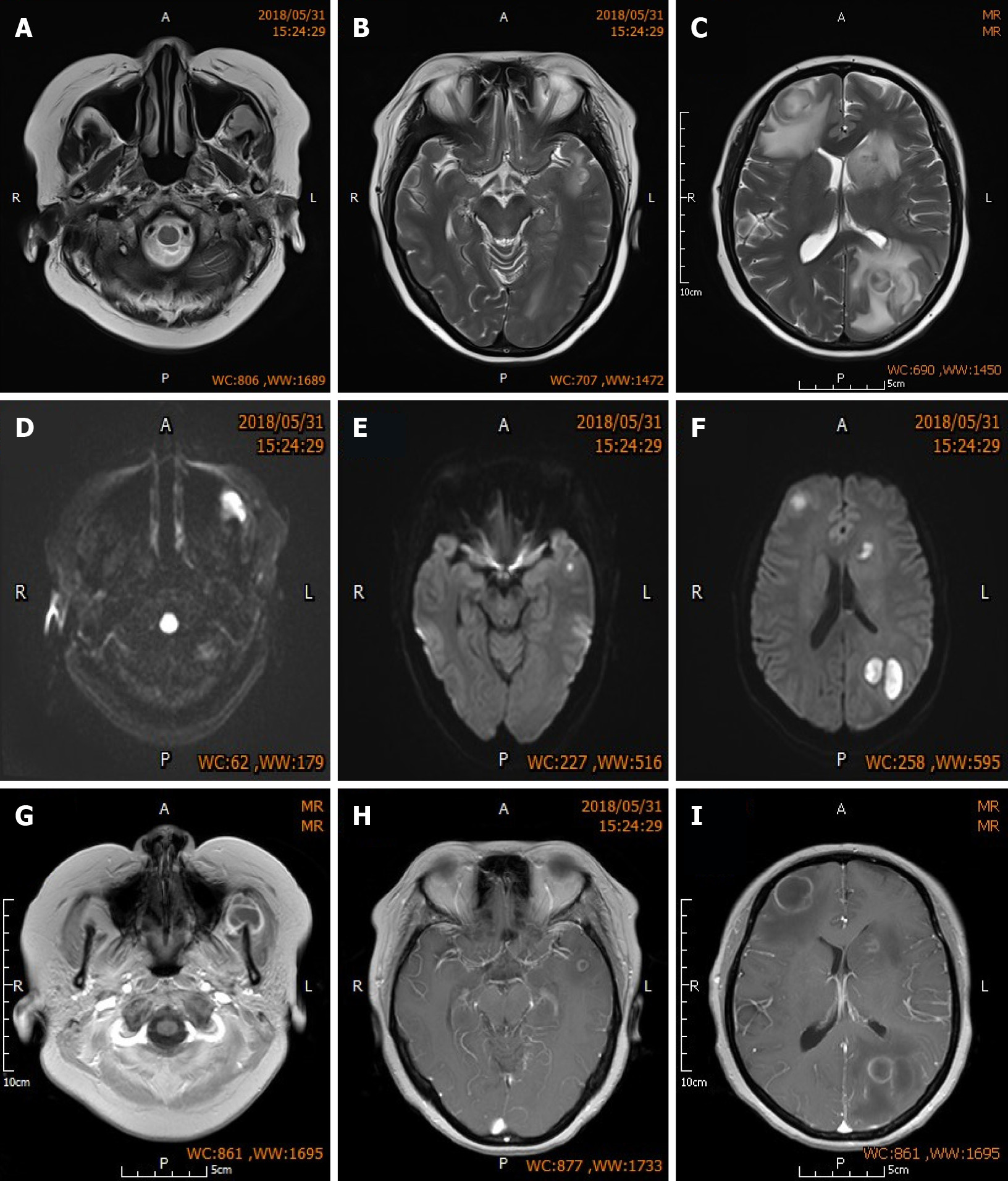
Figure 1 Brain magnetic resonance imaging of the patient.
A-C: Sequential MR-T2WI of both frontal lobes, left parietal lobe, and left masseteric space. Images show multiple nodular lesions with significant perilesional edema; D-F: Sequential MR-DWI show high level of lesion signal change; G-I: Sequential enhanced MR-T1WI showing low lesion signals that are slightly higher than the cerebrospinal fluid signal, and uniform, intact and round annular enhancement around the lesions.
- Citation: Hu QD, Liao LS, Zhang Y, Zhang Q, Liu J. Surgery and antibiotics for the treatment of lupus nephritis with cerebral abscesses: A case report. World J Clin Cases 2022; 10(6): 1981-1990
- URL: https://www.wjgnet.com/2307-8960/full/v10/i6/1981.htm
- DOI: https://dx.doi.org/10.12998/wjcc.v10.i6.1981


