Copyright
©The Author(s) 2022.
World J Clin Cases. Feb 26, 2022; 10(6): 1973-1980
Published online Feb 26, 2022. doi: 10.12998/wjcc.v10.i6.1973
Published online Feb 26, 2022. doi: 10.12998/wjcc.v10.i6.1973
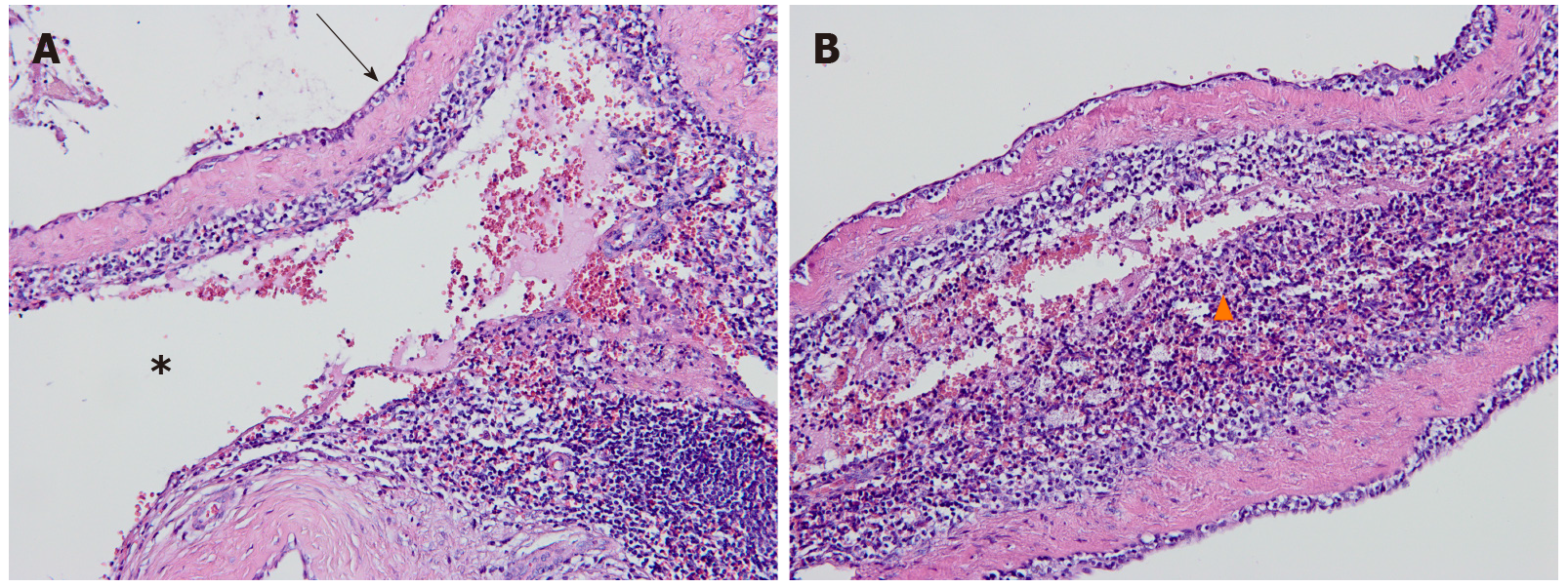
Figure 3 Hematoxylin-eosin staining of the cavernous hemangioma arising from the intrapancreatic accessory spleen.
A: Large dilated vascular spaces (asterisk) separated by fibrous septa and endothelial cells (arrows) lining on the surface of the vascular spaces were observed in the intermediate-power view (original magnification, 200×); B: A high-powered photomicrograph (original magnification, 400×) illustrated splenic tissues (triangles) adjacent to the vascular spaces.
- Citation: Huang JY, Yang R, Li JW, Lu Q, Luo Y. Cavernous hemangioma of an intrapancreatic accessory spleen mimicking a pancreatic tumor: A case report. World J Clin Cases 2022; 10(6): 1973-1980
- URL: https://www.wjgnet.com/2307-8960/full/v10/i6/1973.htm
- DOI: https://dx.doi.org/10.12998/wjcc.v10.i6.1973


