Copyright
©The Author(s) 2022.
World J Clin Cases. Feb 26, 2022; 10(6): 1937-1945
Published online Feb 26, 2022. doi: 10.12998/wjcc.v10.i6.1937
Published online Feb 26, 2022. doi: 10.12998/wjcc.v10.i6.1937
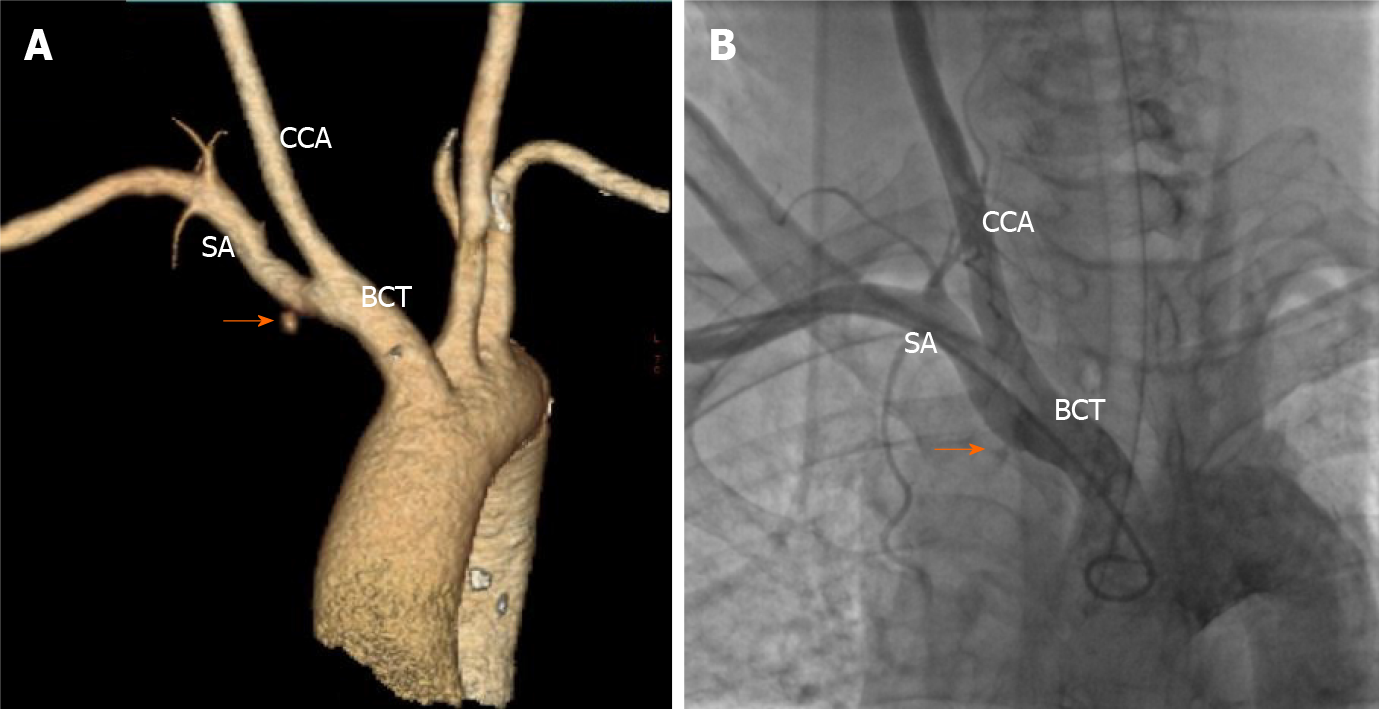
Figure 3 Contrast-enhanced computed tomography and brachiocephalic angiography.
A: Contrast-enhanced computed tomography showing contrast extravasation surrounding the proximal subclavian artery (SA) (orange arrow); B: Brachiocephalic angiography revealing the site of bleeding at the root of the right SA at the intersection of the right common carotid artery (orange arrow). SA: Subclavian artery; CCA: Common carotid artery; BCT: Brachiocephalic trunk.
- Citation: Shi F, Zhang Y, Sun LX, Long S. Life-threatening subclavian artery bleeding following percutaneous coronary intervention with stent implantation: A case report and review of literature. World J Clin Cases 2022; 10(6): 1937-1945
- URL: https://www.wjgnet.com/2307-8960/full/v10/i6/1937.htm
- DOI: https://dx.doi.org/10.12998/wjcc.v10.i6.1937


