Copyright
©The Author(s) 2022.
World J Clin Cases. Feb 16, 2022; 10(5): 1723-1728
Published online Feb 16, 2022. doi: 10.12998/wjcc.v10.i5.1723
Published online Feb 16, 2022. doi: 10.12998/wjcc.v10.i5.1723
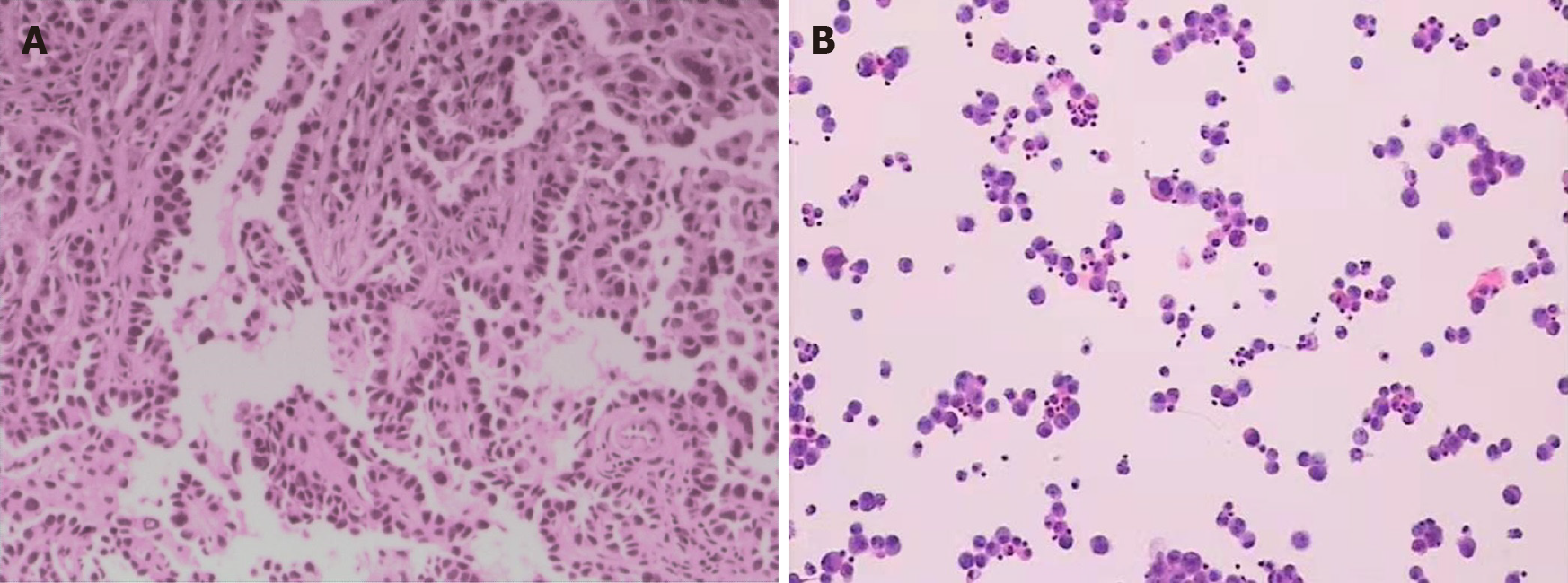
Figure 1 Results of pathologic diagnosis and cerebrospinal fluid cytology.
A: H&E staining, magnification 100×, demonstrated abnormal epithelioid cell nests in the left frontal lesion; B: Cerebrospinal fluid (CSF) cytology revealed malignant cells in the CSF.
- Citation: Li N, Wang YJ, Zhu FM, Deng ST. Unusual magnetic resonance imaging findings of brain and leptomeningeal metastasis in lung adenocarcinoma: A case report. World J Clin Cases 2022; 10(5): 1723-1728
- URL: https://www.wjgnet.com/2307-8960/full/v10/i5/1723.htm
- DOI: https://dx.doi.org/10.12998/wjcc.v10.i5.1723


