Copyright
©The Author(s) 2022.
World J Clin Cases. Feb 16, 2022; 10(5): 1702-1708
Published online Feb 16, 2022. doi: 10.12998/wjcc.v10.i5.1702
Published online Feb 16, 2022. doi: 10.12998/wjcc.v10.i5.1702
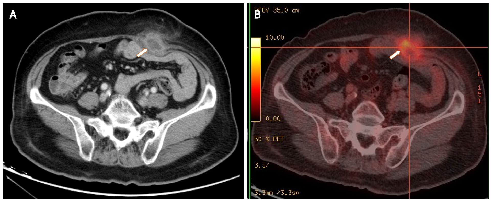
Figure 1 Images of the mass by contrast-enhanced abdominal computed tomography and 18F-fluorodeoxyglucose-positron emission tomography.
A: Computed tomography (CT) shows a low-density mass with rim enhancement adjacent to the rectus abdominis in the lower abdominal wall; B: 18F-fluorodeoxyglucose-positron emission tomography/CT shows high uptake of fluorodeoxyglucose by the mass, with a maximum standardized uptake value of 6.0. The white arrows indicate the mass.
- Citation: Yu Y, Feng YD, Zhang C, Li R, Tian DA, Huang HJ. Aseptic abscess in the abdominal wall accompanied by monoclonal gammopathy simulating the local recurrence of rectal cancer: A case report. World J Clin Cases 2022; 10(5): 1702-1708
- URL: https://www.wjgnet.com/2307-8960/full/v10/i5/1702.htm
- DOI: https://dx.doi.org/10.12998/wjcc.v10.i5.1702


